Thursday, May 30, 2024

Protons (72) Humans: 8% LA vs 74% LA by sustained oral ingestion

This study in humans is very different to the previous rat study. People were fed repeated oral fat loads (you could call this Bulletproof Cocoa rather than Bulletproof Coffee), once an hour for 12 hours then every two hours overnight until the start of an hyerglycaemic clamp at 24h, through which the oral fat loading continued. Glucose was infused to a stable 20mmol/l and insulin allowed to respond as best it might. Insulin sensitivity was determined by the glucose infusion rate in the last 30 minutes of the clamp. In some ways this is more physiological than the hyperinsulinaemic euglycaemic clamp, which is considered the gold standard. The oral fat ingestion was slightly above calculated 24h caloric requirements for these subjects.

Differential effects of monounsaturated, polyunsaturated and saturated fat ingestion on glucose-stimulated insulin secretion, sensitivity and clearance in overweight and obese, non-diabetic humans

Over a 24h period the ingestion of safflower oil, with linoleic acid providing in the region of 70% of total calories, ought to demonstrate the initial Protons predicted insulin sensitising effect, which would only be later replaced by the uncoupling effect if the study had been continued for a week or two. 

Again we can assess insulin sensitivity by how much glucose was needed to be infused during the last 30 minutes of the clamp to maintain an hyperglycaemia of 20mmol/l. This is what happens:


I don't think I have to make any qualifications here. SFA oral ingestion for 24h causes a very similar degree of insulin resistance to oral ingestion of a minimal calorie supplying control chocolate drink. Tallow rather than palm oil would have accentuated the effect.

Ingesting 70% of your calories as linoleic acid over a 24h period is insulin sensitising compared to ingesting SFA, p less than 0.001. Or ingesting virtually nothing at all, p < 0.05.

Linoelic acid is insulin sensitising.

This is BAD. When fasting you *must* resist even basal insulin or that insulin will lower fasting glucose, lower fasting FFAs and you will be hungry. And raid the fridge at 2am. And get fat.

Protons.

Peter

Protons (71) Rats: 13% LA vs 61% (mostly) LA by infusion

I have a certain, very specific, idea of how linoleic acid produces obesity. It seems as though relatively few people share this point of view. That is absolutely fine. Bright people have their own ideas and, eventually, if the core process is consistent, all views of the development of obesity and its associated insulin resistance will eventually converge. I spend a great deal of time thinking about whether linoleic acid enhances insulin sensitivity -> directly causing obesity or whether linoleic acid causes insulin resistance directly -> reactive hyperinsulinaemia -> obesity. The data make me favour the former.

This is the first paper I have come across where various fatty acid mixtures were assessed, in rats, for their acute effects on insulin sensitivity in vivo. In particular I was interested in the effect on glucose utilisation under hyperinsulinaemic euglycaemic clamp conditions. The higher the infusion rate, the more insulin sensitive the rat is.

They infused intravenous oil emulsions continuously for five hours and then continued throughout the exogenous hyperinsulinaemia over the following two hours, while clamping glucose at around 6.5mmol/l. So this is looking at normoglycaemia combined with fasting levels of FFAs until the clamp period. The rate of deliver per hour was roughly comparable to a 24h intake of calories for a rat of this size, averaged to an hourly rate.

Everything is fairly physiological until you add in the insulin/glucose infusions for the clamp while maintaining the lipid supply. Then you are looking at the situation where FFA supply cannot be suppressed by insulin, so you have a model for metabolic syndrome.

The results are quite clear. Whole body insulin responsiveness is suppressed by any fatty acid availability.


Clearly the glucose infusion rate, representing whole body insulin sensitivity, is lower in the SATU group (lard oil) compared to the PUFA group (soybean oil) but this is not remotely statistically significant (p = 0.2849). However there is no suggestion that linoleic acid is uniquely triggering insulin resistance compared to saturated fats, bearing in mind that modern (2015) Canadian lard is higher in insulin sensitising LA at 15% than my preferred fats such as beef tallow or suet which are around 2% LA (correctly ignoring any CLA content).

So the Protons concept could be suggested to have earned some marks here, there is more insulin resistance in the saturated fat group (GIR 43micromol/kg/min) when compared to the less insulin resistant linoleic acid infused group (GIR 73 micromol/kg/min). But not statistically significant.

However, there is no suggestion that linoleic acid per se causes enhanced insulin resistance, so causing obesity via secondary hyperinsulinaemia. In fact the trend is in the reverse direction.

In these rats.

Humans next.

Peter

Wednesday, May 29, 2024

Foie Gras (11) Hepatocyte mitochondria


Another tidy up, this time related to 

Fat Quality Influences the Obesogenic Effect of High Fat Diets

and the paradox of mitochondrial uncoupling in section B of Fig 4:


It is patently obvious from this plot that mitochondria extracted from the liver tissue of lard fed rats (consuming an obesogenic level of linoleic acid) do have an higher uncoupled oxygen consumption at all values of membrane potential when compared to the level of oxygen consumption in those rats fed the high safflower oil diet.

That is exciting and paradoxical.

We know from Figure 1 that the safflower oil fed rats were more uncoupled overall than the lard fed rats. They were synthesising much more UCP-1/cell in their brown adipose tissue and they had a greater absolute mass of brown adipose tissue by the end of the study.


They were also actively expending more energy per day at the end of the study compared to the lard fed rats. This is stated in the legend to Figure 2:

"Percent contribution of lipids, proteins and carbohydrates to total daily energy expenditure (lard = 380 ± 15, safflower-linseed = 410 ± 25 kJ/day x kg0.75) in rats fed lard or safflower-linseed high fat diet."

I would expect all rats/mice fed high safflower oil diets to use this technique and so eventually normalise their weight to that of chow fed rats/mice on a long term basis, as was found (in mice) here:

Prevention of diet-induced obesity by safflower oil: insights at the levels of PPARalpha, orexin, and ghrelin gene expression of adipocytes in mice

Okay, let's summarise:

Safflower oil induced an initial obesity by increasing insulin sensitivity which was, by day 14, in the process of being reversed by UCP-1 reducing that excessive insulin sensitivity in WAT, assisted by activating BAT.

No one would expect hepatocytes to express UCP-1, they just don't do this. The liver deals with excess calories by sequestering them as triglycerides under the influence of insulin, sequestering them as triglycerides under the influence of succinate derived from peroxisomal omega oxidation or by signalling to BAT using FGF21 as a mediator to increase UCP-1 expression so as to bulk off-load calories as heat. But not in the liver.

Soooooo.

Safflower oil (~70% linoleic acid) produces whole-body uncoupling in the rats in the current study, apparently with the exception of within liver tissue.

Hepatocytes *do* use UCPs, they definitely synthesise UCP-2 and UCP3, but not for bulk lipid oxidation. Current thinking is they are used to fine tune their inner mitochondrial membrane potential while other signals deal with bulk caloric overload.

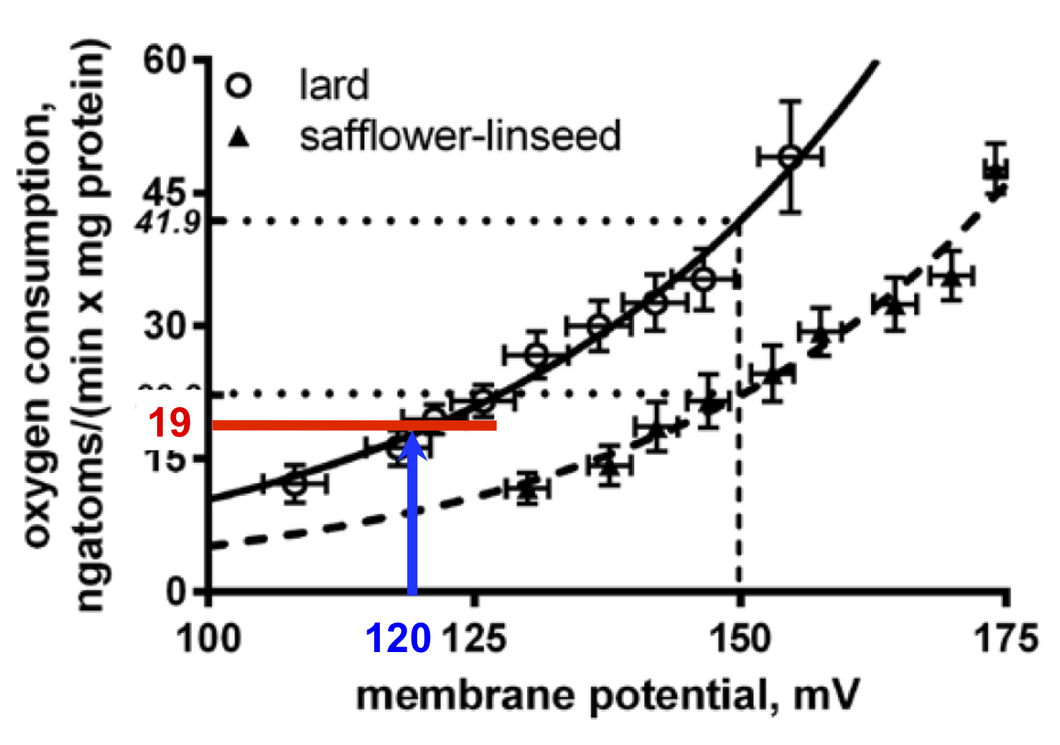
So the paradox is that lard fed rats have more uncoupled mitochondria than safflower fed rats. That's what the graph at the top of the page shows, ie hepatocytes are doing the opposite of what the whole rat is doing...

They measured delta psi of isolated mitochondria with a dye (safranin O) calibrated back (through 4 layers of references) to the standard technique which gives us our best estimation (don't ask) of membrane potential. They then fed isolated mitochondria in the presence of oligomycin (to block ATP synthesis) and rotenone (to prevent RET through complex I). At this point all oxygen consumption is from uncoupling. If you add increments of malonate to progressively inhibit complex II you can progressively lower the delta psi and look at the degree of uncoupling at a given titrated delta psi.

On the face of it it looks very much as if the liver really is doing the opposite to the rest of the body, which seems counter intuitive:


The degree of uncoupling is being assessed at a fixed potential, here the group chose to use 150mV (the blue line) for their example, giving an uncoupled oxygen consumption of 41.9 in lard fed vs 22.2ngatoms/(min x mg protein) of oxygen if safflower oil fed.

But is this the case in vivo? The lard fed rats are chronically underfed and have lipid locked in to adipocytes by excessive insulin sensitivity so what little lipid is being released is via augmented basal lipolysis. It is completely plausible (but also completely made-up) that they might be running a membrane potential, in vivo, as low as 120mV. Like this, blue line:


At 120mV you are not going to making a lot of ATP so uncoupling would be actively disadvantageous. In this example the uncoupled oxygen consumption would be low, in the region of 19ngatoms/(min x mg protein) of oxygen, red line.

The safflower oil fed rats went through an initial hypocaloric episode during their initial weight gain phase, but now they are uncoupling in WAT which will blunt insulin signalling and release a surfeit of FFAs, enough to supply large amounts of FFAs the liver mitochondria and (in parallel) accumulate as lipid droplets to the point of cellular damage occurring.

Under this level of direct hepatic caloric excess the mitochondrial membrane potential is likely to be high. If we run another thought experiment (ie make up) a potential of 160mV, just under that 170mV threshold for marked ROS generation, this would give us an uncoupled oxygen consumption of 29ngatoms/(min x mg protein) like this:


So, if the membrane potential differs between groups in vivo, so would the level of uncoupling. It is completely plausible that (safflower) lipid overloaded mitochondria are running an high delta psi, so need more uncoupling. Mitochondrial will never have a fixed delta psi of 150mV. It is absolutely possible that, in vivo, the safflower oil fed rats had more uncoupled hepatic mitochondria compared to the lard fed rats.

I feel much more comfortable with having hepatocytes uncouple *more* with safflower oil than with lard. The whole study is bias confirming of multiple aspects of the Protons hypothesis. Things have to make sense.

I have no problem with the mitochondrial preparation the group developed here and how they have used it. It's no better/worse than any other mitochondrial preparation. What is crucial is how you interpret the data it provides you with in the light of what must be happening physiologically. Then extrapolate backwards to the most plausible in-vivo situations, with caveats.

I have my biases.

Peter

Late addendum.

The mitochondrial uncoupling curve I have been discussing was generated from mitochondria treated with FFAs to facilitate uncoupling. Of course, if you argue that the lard fed, hypocaloric rats had lower levels of FFA in the fed state in vivo (in the fasted state there is no difference in FFA level) then there would be much less uncoupling than that discussed above, emphasising the point. Without FFA supplementation, at 160mV in the lard fed rats uncoupled oxygen consumption was as low as ~5ngatoms/(min x mg protein) in section A of Figure 4. In reality it would be somewhere between this value and the FFA supplemented value. As much as any mitochondrial prep reflects reality.

Foie Gras (10) Liver

Just to tidy up my thoughts on


TLDR: I suppose all I really have to say is that the title is incorrect and the scrutineers are completely incompetent.

We have these data for mRNA production from "pro-inflammatory" genes in liver tissue:


which we know, from their section of adipose tissue, have absolutely zero correlation with active inflammation, which they assessed in adipose tissue using the activity of the myeloperoxidase system.

The liver is full of resident macrophages, known as Kuffper cells, which are very good at activating their myeloperoxidase system. The group has an assay for this activity. They didn't use it on liver tissue. Why not?

I have no idea whether the liver macrophages actually used any of the reported mRNA products to generate inflammation. 

Go figure.

The group did, for some reason, measure plasma CRP levels, CRP being an acute phase protein produced by the liver in response to any inflammation, *anywhere* in the body. It might have been raised in response to the activity of the myeloperoxidase in the adipose tissue of the HF fed mice, that might be logical. We'll never know because they omitted to measure CRP in the plasma of the HF fed mice.

Go figure. I have not edited this chart in any way:


So we know essentially nothing about inflammatory changes in the liver and we know nothing about the levels of CRP produced (or not) in the plasma of HF diet fed mice. Which did have inflamed adipose tissue and *might* have had inflamed liver tissue.

Oh, their one interesting finding was that saturated fat is suppressive of "pro-inflammatory" gene expression in liver tissue. But not in adipose tissue.

Would this be protective against inflammatory liver damage? There is no way you can assess this from this study, but the idea is nice.

But ultimately the liver section of the paper is complete dross.

I said it before, these people are rank amateurs.

Peter