Thursday, June 23, 2022

FFA vs 4-HNE for activating uncoupling

Another basic mitochondrial concept. This is Brand again. The paper features mitochondria extracted from yeast cells which have been transfected with a plasmid for the mammalian UCP-1 gene.

Synergy of fatty acid and reactive alkenal activation of proton conductance through uncoupling protein 1 in mitochondria

UCP-1 is the odd man out of UCPs, its primary function is thermogenesis in adipose tissue but it seems that the control systems are similar across the whole family of proteins. UCP-1 is useful because the degree of proton leak is huge compared to other UCPs, which makes measurements using isolated mitochondrial preparations easier.

It turns out that, in addition to palmitate (and other fatty acids) many lipid derivatives also activate uncoupling, 4-HNE being one of the best studied.

They isolated mitochondria from their yeasts and fed them with either 4-HNE, palmitate or a combination of the two and looked at the degree of uncoupling (using O2 consumption under oligomycin as the surrogate, as you do).


The two red rectangles are the degree of uncoupling induced by either 4-HNE alone or palmitate alone. Both do something. If you simply add the two red rectangles together you get the blue one, which is what you would expect if the two agents were additive. The yellow rectangle is what you actually do get, ie significantly more uncoupling because the combination is synergistic. There are papers which suggest the 4-HNE is essential for palmitate to uncouple but that might be model dependent. The above experiments are using a membrane potential of 87mV, ie quite low. A high membrane potential might have generated enough 4-HNE in situ to mask the effect of exogenous 4-HNE. Or done other things, next post.

Philosophically I view the 4-HNE in this role as a signal that some degree of ROS related "damage" has occurred to the PUFA components of the mitochondrial membrane. A little too much in the way of ROS produces a degree of lipid damage which facilitates uncoupling, which drops mitochondrial membrane potential and lowers ROS generation and subsequent damage. I have the quotation marks around "damage" because the degree of damage is that at which evolution has decided is acceptable before stepping in with an effective intervention, ie the damage is permissible and non injurious to the cell.

Bottom line: Fatty acids and lipid oxidative derivatives of PUFA both support uncoupling. Their mechanisms appear to be different and to be synergistic. We can go on to look at some aspects of their regulation in the next post.

Peter

Saturday, June 18, 2022

Insulin increases coupling in mitochondria

Back in the 1990s Veech's lab noted that supra maximal insulin, combined with glucose at 11mmol/l, markedly improved the ability of an isolated rat heart to pump oxygenated perfusion fluid compared with glucose alone. The mechanism of the effect was not explicable from their model but was very clear cut and the time scale of onset suggested a covalent bonding process.

Substrate signaling by insulin: a ketone bodies ratio mimics insulin action in heart

Macroscopically, the amount of work done per mole of oxygen consumed increased. As this was without an increase in glycolysis the implication is that insulin increases the coupling of mitochondria.

I was left with the idea at the time, reinforced occasionally by other finds, that insulin was has a major effect of increasing coupling within mitochondria.

Insulin clearly has many, many effects within a cell. It's not possible to examine any of these using isolated mitochondrial preparations because they have no cytoplasm to respond to insulin. You need intact cells.

I recently came across this rather nice paper:

Insulin acutely improves mitochondrial function of rat and human skeletal muscle by increasing coupling efficiency of oxidative phosphorylation

It looks at an assortment of muscle derived cells in much the same way as mitochondrial preparations examine mitochondrial performance, but here whole cells used, a small step closer to reality than isolated mitochondria. They have intact cytoplasm so can function on "normal" substrates such as glucose or palmitic acid. The cells are not even "permeabilised".

They used standard mitochondrial techniques such as full uncoupling with FCCP to assess the maximum possible oxygen consumption and oligomycin to assess peak oxygen consumption from proton leak in the absence of a functional ATP synthase. So they can provide standard mitochondrial study parameters like respiratory control ratio and make estimates of the degree of (un)coupling of respiration and of the efficiency of ATP generation.

All good but even better they then went on to look at the effect of fairly physiological concentrations of insulin on these parameters. And to look at the effect of palmitate alone and palmitate in combination with insulin.

They are looking at mitochondrial function within intact cells, with functional cell surface receptors and cytoplasmic signalling cascades. Insulin was used at 10nmol/l (10,000pmol/l) which is only just above peak post prandial levels and even their 100nmol/l dose is still way below the millimolar concentrations commonly used to assess the effects of supra maximal insulin stimulation on cell preparations.

Their palmitate dose rate is hard to assess as they presented it bound to albumin with an estimated free palmitate of 20nmol/l, ie 0.02micromol/l. Almost every other study simply measures/specifies total palmitate in solution so making comparisons is hard. Obviously the 400-2000micromol/l of FFAs which are normal in fasted human plasma are almost completely albumin bound, so it's hard to tell if the estimated 20nmol/l of free palmitate used in the study is high or low. It certainly has an effect.

These are the graphs of oxygen consumption from the human derived muscle cells/myotubes:


The graphs are not intuitive. First, everything is normalised to the rate of oxygen consumption under the influence of oligomycin (between times 20 and 40 minutes) ie state 4oligomycin, and are expressed as a percentage of this. So the sections of the graph in the "dip" after the line labelled "OLI" are baseline and labelled 100, ie 100%.

Under oligomycin there is a complete blockade of ATP synthase so any oxygen consumption has to be facilitated by uncoupling. The absolute values will not be identical with vs without insulin, they are just deliberately aligned at 100. The absolute values will differ based on the activity of uncoupling proteins.

Once FCCP is added there is complete uncoupling of all respiration (while ATP synthase still remains blocked with oligomycin) and so this represents the maximum possible flow of electrons down the ETC to complex IV, with no buildup of proton gradient to inhibit this. These peak values are probably identical whether insulin has or hadn't been applied because FCCP is supra maximal in its uncoupling so subtleties of UCPs become irrelevant. No one has added palmitoylcarnitine either.

This shows as a greater percentage *increase* when insulin has been applied earlier, ie the oligomycin phase had different absolute oxygen consumptions with or without insulin. I think it's just convention to set up the graphs as they are.

So glucose + insulin couples respiration compared to glucose without insulin.

Which reiterates Veech's findings.

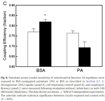
Okay. So we can calculate the coupling efficiency of mitochondria respiring on glucose with or w/o insulin and express it as a fraction of unity. Insulin always increases the coupling of respiration when oxidising glucose. Black bars with insulin:


The study didn't look at delta psi or ROS generation so we have no way of knowing exactly what happens to these parameters.

Adding palmitate completely blocks (and probably (ns) decreases) the increase in respiratory coupling seen when insulin is added to glucose. Right hand columns labelled as added PA:


The change downward looks to have come very close to statistical significance. This suggest that small (possibly) doses of palmitate negate insulin's coupling effect and trend towards actively reversing it.

What is also interesting is the left hand pair of bars. The white bar is insulin + glucose and the black bar is insulin, glucose and "empty" bovine serum albumin (BSA). The BSA produces a statistically significant increase in coupling of respiration. This is in a cell prep which has not been treated with exogenous fatty acids. There are enough fatty acids "floating around" to interfere with insulin's coupling action on mitochondria.

My assumption is that the empty BSA scavenges free fatty acids by supplying a sequestration site for any FFAs in the culture. Reminiscent of the effect of carnitine in a previous post.

Let's make this completely clear: Mitochondria in cells exposed to insulin are more coupled compared to those without insulin. Adding extra palmitic acid reduces this extra coupling. Removing background levels of free fatty acids enhances insulin's coupling effect.

Insulin is an enhancer of coupling in the mitochondria of intact cells. It's effect appears to be mediated through changes in free fatty acid availability which are known mediators of activation of uncoupling proteins.

TLDR: All isolated mitochondrial preparations are devoid of insulin signalling so will automatically be uncoupled to some degree, which goes some way to explaining continued oxygen consumption under oligomycin. Especially using supra maximal NADH generating substrates. But it doesn't help explain the regulation of membrane potential to around 180mV under high substrate supply.

Other things might.

Peter

Monday, June 06, 2022

Beta oxidation intermediates control ETC function

Back to the paper provided by met4health, briefly mentioned previously:

Electron Transport Chain-dependent and -independent Mechanisms of Mitochondrial H2O2 Emission during Long-chain Fatty Acid Oxidation

It's a very interesting paper. It brings to light some of the problems of using isolated mitochondrial preparations. The basic summary is that if you compare the oxidation of palmitoylcarnitine to either pyruvate/malate or glutamate/malate there is significant ROS generation with palmitate, even at low delta psi, compared to the primarily NADH generating substrates (which produce zero ROS at 180mV delta psi in these preparations).

Figure 3 is perhaps the most interesting. For section C they blocked the function of ATP synthase with oligomycin to raise delta psi and then titrated delta psi downwards by uncoupling with FCCP to give either a low or high delta psi. Then they fed the preparation with either palmitoylcarnitine (plus extra carnitine) or glutamate/malate.


The right hand bar graph is derived from the left hand curve and shows that delta psi has some influence but, under both delta psi conditions, there are many more ROS produced under palmitate oxidation than G/M. Delta psi clearly has an effect but substrate also has a marked influence.

That's the convincing part of the paper. There is no insight as to mechanism but my biases assume it will be F:N ratio related. I might have left it there but I can't.

The rest of the results are hugely influenced by this statement from the methods:

"Unless otherwise stated, determinations were made in the presence of oligomycin (3µg/ml) to inhibit ATP synthesis, a condition used in previous studies of the mechanisms of ROS formation in isolated mitochondria and permeabilized muscle fibers (e.g. see Refs. 4, 13, 34, and 38)."

Translation: We blocked ATP synthase because everyone does it.

So pretty well all of the results appear to have been produced during a complete blockade of ATP synthase.

These preparations are always looking at ROS generation when the only dissipation route for membrane potential is some form of uncoupling (UCPs, NNT, various proton assisted co-transporters)

There is no other way to allow O2 consumption under oligomycin. See last post on Brand's review.

This begs the question of how is it possible to feed a mitochondrial preparation supra maximal amounts of NADH substrates (pyruvate/malate) consuming relatively large amounts of oxygen in the absence of any way of dissipating the proton gradient across the inner mitochondrial membrane without ATP synthase being active?

The answer is that there must be some sort of uncoupling going on. The delta psi of 180mV is completely normal but this does not mean that there is no proton leak through the inner mitochondrial membrane. It merely means that the leak (defined by an O2 consumption of 21nmol O/min/mg of mitochondrial protein) is sufficient to avoid raising delta psi to massive levels in the face of a supra maximal supply of pyruvate/malate. The proton leak is also kept low enough not to drop delta psi. This smacks of regulation.

By comparison palmitoylcarnitine at 18micromol/l has less oxygen consumption and supports a lower delta psi, in the region of 145mV.

This is clear in section A of Figure 3.


Both of these values for O2 consumption under oligomycin *have* to be facilitated via uncoupling.

What is also clear is that, with oxygen consumption lower under palmitoylcarnitine, there is less uncoupling than for P/M. 

Has anyone noticed that in Figure 3 there is a flick between 18µM palmitoylcarnitine and 18µM palmitoylcarnitine plus 2mM carnitine between various graphs?

The authors of the paper considered that, with palmitoylcarnitine, the low delta psi combined with low O2 consumption might be due to an inhibitory effect of fatty acid oxidation intermediates on either FAO itself or on ETC function.

Adding 2mM carnitine appears to remove such intermediates. I've not been in to the chemistry but it looks like the carnitine exports them from the mitochondria. There's something about this in the supplementary data. I'm just accepting it happens for today. About which I'm a little cautious.

Adding the extra carnitine makes the oxidation of palmitoylcarnitine look just like supra maximal P/M or G/M.

Here's the oxygen consumption bar chart from supplementary data Figure 3. We're looking at the left hand pair. White bar is palmitoylcarnitine 18µM consuming (as before) 10nmol O/min/mg. Black bar is after the extra carnitine was added. Oxygen consumption is around 25nmol O/min/mg (and delta psi did the same) and is now comparable across the metabolic substrates, black bars:


This increase in O2 consumption means that, under oligomycin, that uncoupling has markedly increased and is directly equivalent to supra maximal NADH sources.

To me this implies that normal fatty acid oxidation (ie without extra carnitine) is a self limiting process. 

If the paper is correct (don't forget they are working with oligomycin blocked preparations) a high delta psi is not a feature of palmitoylcarnitine oxidation.  The oxidation of palmitoylcarnitine suports ROS generation irrespective of delta psi. To make me really happy it would be nice to generate ROS with different fatty acids and look at the effect of the F:N ratio on ROS generation.

There are certain implications to these thoughts. First is that physiology is very keen to keep delta psi in the region of 180mV or lower and applies some degree of uncoupling to achieve this. This appears to be independent of fatty acid induced uncoupling.

This is possibly very important. Any supra maximal supply of substrate in a non-phosphorylating mitochondrial prep (state 4oligomycin) has to have a method to stabilise delta psi at around 180mV. How? Another post there.

Second is that FAO intermediates down regulate the ETC performance directly, limiting delta psi to 145mV from palmitoylcarnitine 18µM unless those FAO intermediates are removed.

Finally, FAO appears to generate ROS moderately independently of delta psi. Certainly palmitoylcarnitine does.


Now for a long-time-ago throwback:

Does everyone recall the Dutch chaps who didn't eat for 60 hours and so rendered their mitochondria "dysfunctional"?

Prolonged fasting identifies skeletal muscle mitochondrial dysfunction as consequence rather than cause of human insulin resistance

Permeablised  muscle fibres behave pretty much like mitochondrial preparations.

"Despite an increase in whole-body fat oxidation, we observed an overall reduction in both coupled state 3 respiration and maximally uncoupled [here using FCCP] respiration in permeabilized skeletal muscle fibers..."

The RCR using an uncoupler and oligomycin, ie state 3FCCP / state 4oligomycin, fell markedly with fasting, hence the term "skeletal muscle mitochondrial dysfunction" in the title.

But 60 hours of fasting cannot possibly destroy your mitochondria. People can pushbike hundreds of kilometres over 5 days without eating anything at all. Their mitochondria work.

I would suspect that this is a fully physiological control system designed to cope, at the mitochondrial level of fatty acid oxidation, with a potentially limitless supply of energy from the fatty acids released from adipocytes under low insulin/insulin signalling conditions.

In this study using permeablised muscle fibres you could probably have reversed the effect completely by treating with 2mM carnitine.

But why would you want to? Apart from gaining insight as to what is normal physiology of course.

Summary: fatty acid oxidation is a self regulating system at the level of beta oxidation rather than at the level of the Krebs Cycle. I suspect that the FAO intermediates will act directly on the electron transport chain.

If this is correct it will provide insight in to other elevated FAO conditions, ie obesity with insulin resistance, where fatty acids should modify (appropriately) ETC function to avoid energetic overload.

I've had suspicions that this has to be the case for a long time. Finally I'm getting to see a little progress.

Peter

Brand insights

In the comments to a previous post met4health provided a link to this paper:

Electron Transport Chain-dependent and -independent Mechanisms of Mitochondrial H2O2 Emission during Long-chain Fatty Acid Oxidation

Before we can go in to it in depth we have to define a few terms. For this we need Brand's excellent review

Assessing mitochondrial dysfunction in cells

which clarifies many of the terms related to mitochondrial function and their variations between experimental set ups.

Perhaps first term we should look at is the Respiratory Control Ratio (RCR). If you feed a quiescent isolated mitochondrial preparation with substrate but no ADP it consumes a small amount of oxygen. If the preparation is given a briefly supramaximal concentration of ADP (in the presence of permanently high phosphate) there will be a marked rise in O2 consumption while the ADP is converted to ATP. This peak O2 consumption represents the maximum respiration under relatively "physiological" conditions. This is state 3 respiration.

If left alone the ADP is fairly quickly used up and O2 consumption will drop back to basal levels. This level is state 4 respiration.

The RCR is state 3 (maximum possible O2 consumption) divided by state 4 (basal O2 consumption). Conceptually this is a marker of how good a mitochondrial preparation is at upping ATP production when needed. High RCR suggests excellent function.

People have modified the routes to these numbers. The first modification is converting the "idling" state 4 to a "stationary" state. This is done with oligomycin, a complete ATP synthase inhibitor. There are reasons this is done but for now we can just accept it. Brand uses the term state 4oligomycin or state 4o.

Next modification, instead of looking at maximum O2 consumption under surplus ADP, is to simply use a chemical uncoupler to probe maximum possible O2 consumption. This is the peak value of state 3 or state 3 uncoupled (state 3u). If you happen to have used FCCP as your uncoupler you might use the term state 3FCCP.

So RCR is simplified to O2 consumption under FCCP divided by O2 consumption under oligomycin. Crude but effective.

Also Brand says this:

"Net forward flux through each electron transport complex requires a thermodynamic disequilibrium, i.e. the free energy available from electron transfer must be greater than that required to pump protons across the membrane against the pmf."

This translates as: if the proton motive force (pmf) is very high then zero electrons will travel down the ETC, none will reach complex IV and zero O2 will be consumed.

Think about that. Without dissipating a high pmf there is no O2 consumption. This suggests that O2 consumption in state 4oligomycin must be synonymous with uncoupling of various types to allow any O2 consumption at all (necessitating pmf dissipation) in the absence of a functional ATP synthase.

This lets you qualify the information provided by papers which quantify the O2 consumption under oligomycin without mentioning the significance of oxygen being consumed in the absence of a functional ATP synthase.

Brand's review is full of such gems as the above but I think these insights are enough to allow us to go on and look at ROS generation from fatty acid oxidation in the presence/absence of elevated pmf (the electrical component of which is delta psi).

Peter