Wednesday, March 29, 2023

Metformin (13) PowerPoint doodles

Sorry if this is a bit repetitive, I have a paper on metformin I'd like to discuss and just to make sure I understand how metformin works I've re-read this paper from many moons ago, just to be sure I'm clear 

Metformin suppresses gluconeogenesis by inhibiting mitochondrial glycerophosphate dehydrogenase

So here we go with PowerPoint once more:

This the route for glucose production starting from glycerol


which is very straight forward. Let's flesh it out with some enzymes. For gluconeogensis we need to convert glycerol-3-P in to DHAP, this is performed by mitochondrial glycerophosphate dehydrogenase thus:


Of course this is half of the glycerophosphate shuttle, the other half being thus:


and we can put the two together to show the full shuttle


and then we can inhibit mtG3Pdh using metformin


and gluconeogensis grinds to a halt. Glycerol-3-P and NADH accumulate in the cytoplasm. If cytG3Pdh tries to run in reverse (which it can) it simply forces the accumulation of NADH in the cytoplasm, to which there are limitations. There's also a secondary change as regards lactate.

Here's how lactate is used, as it usually is, for gluconeogenesis. 


using lactate dehydrogenase and NAD+ to generate the pyruvate


so of course an high NADH level under metformin will make this process impossible, even if the pyruvate could later get past the inhibited glycerophosphate shuttle. Rising NADH in cytoplasm stops the conversion of lactate to pyruvate. Lactate accumulates.


That's it. Metformin suppresses hepatic glucose output by inhibiting gluconeogensis at the mtG3Pdh step. It acts it within minutes of an IV bolus in an alive rat at therapeutic plasma concentrations. Lactate accumulation is secondary to the redox changes in the cytoplasm.

Let's rephrase that: metformin blockades mtG3Pdh to produce an effect which imitates the suppression of hepatic glucose output by insulin.

This has absolutely nothing to do with enhanced insulin signalling. At all.

Hold on to that.

Peter

Sunday, March 26, 2023

Endocannabinoids in brain and adipocytes


This is very clever stuff. It is possible to engineer in to mice the ability to induce permanent knock-out for the CB1 receptor gene, only in adipocytes and only after the whole mouse is treated with tamoxifen for a few days. So you can grow mice which are phenotypically normal on either chow or an high fat diet (circa 6% LA plus unspecified carbohydrate source) until they are 16 weeks into the study then permanently delete, using tamoxifen exposure, all CB1 receptors on all adipocytes.

Okay, so what happens to food intake if you delete the adipocyte CB1 receptor? It's interesting because some of us (myself included) feel that pathological insulin sensitivity in adipocytes is dominant over brain physiology in terms of weight gain/loss. This paper sums up isolated adipocytes and CB1 activation: 

The CB1 endocannabinoid system modulates adipocyte insulin sensitivity

Without endocannabinoid facilitation of insulin sensitivity you should stop losing calories in to adipocyte fat stores, so you should stop being hungry...

Nothing happens.

Upper line is mice fed on an high fat diet for 16 weeks and then tamoxifen treated to eliminate the adipocyte CB1 receptors in those with the tamoxifen sensitive knockout mechanism. This graph is an amalgamation of sections B and D of Figure 1. I've pulled and stretched both vertical and horizontal scales so they now match for calories and for weeks to make clear what happened. Tamoxifen exposure is circled:


I think we can conclusively say that when adipocytes can no longer listen to the insulin sensitising effects of CB1 receptor activation there is no fall in food intake. Which supports the idea that

CB1 activation -> brain -> eat

so you could say:

↑ dietary LA -> ↑ endocannabinoids -> brain -> eat -> get fat

Nice and simple. It's almost as if appetite regulation by the endocannabinoid system might be brain-centric and adipocytes might be unimportant.

Except. Here's what happens to the weights above baseline. Pulled and stretched and tidied from sections C and E of Figure 1, tamoxifen exposure circled again, as above:


So, from week 17 to week 26 the high fat fed adipocyte CB1 receptor knockout mice continued to eat as much as the high fat fed wild type mice but dropped their weight to match that of the control group.

Without dropping their food intake or linoleic acid exposure. They eat as much junk food as they like and lose weight...

Obviously they uncouple. Not just in brown adipose tissue, white adipose tissue beiges too.

Now: You could simply conclude that brain CB1 receptor activation makes you continue to eat extra and adipose tissue, now without CB1 receptor activation, becomes a calorie sink via uncoupling to dispose of those excess calories eaten under brain CB1 activation. Metabolism is hypercaloric, adipocytes off-load those excess calories. Brain first.

Or: You could conclude that when adipocytes lose their CB1 receptor with its insulin sensitising effect they become less able to store calories and, on an individual cell basis, decide to oxidise their suddenly available stored lipid. If adipocyte hypertrophy includes a lot of linoleic acid then this LA is released and it acts as the best bulk facilitator (along with some 4-HNE) of uncoupling protein activation available. So loss of calories occurs within adipocytes through uncoupling protein activation. Basal lipolysis falls with decreasing lipid droplet size and the brain senses a loss of systemically available calories so maintains food intake to maintain energy homeostasis, ie the adipocytes still control energy availability which controls the brain's action. Metabolism is hypocaloric necessitating food intake. Adipocytes first.

I was going to stop at this point and I probably should have but here's some more rambling anyway.


It has just occurred to me that we can make this comparison:

Mixed diet + LA -> ↑ insulin signal in adipocytes -> loss of calories in to fat droplets -> obesity -> obesity being due to calorie loss in to physical triglyceride storage in adipocytes -> need to eat more (brain sensing ↓ systemic available calories). Getting fat makes you hungry. Fundamental.

This is my standard view of obesity, first pointed out (in my case) via Gary Taubes, although the LA component comes from Protons.

Now we can view the situation under adipocyte CB1 receptor knockout as:

↓ insulin action in adipocytes via ↓ CB1 receptor -> ↑ lipolysis -> ↑ UCP activation -> ↑ loss of fat, but still within adipocytes, only now as CO2 and H2O eventually excreted by lungs/kidneys rather than being released as "calorie carrying FFAs" -> ↓ energy delivery to circulation -> need to eat more (brain sensing ↓ systemic available calories).


Calorie loss in to storage and calorie loss in to uncoupling look the same to the brain.

One makes you fat, the other makes you hot.


Obviously today we already have research drugs to block peripheral CB1 receptors which don't cross to the brain so don't cause the suicidal ideation that central CB1 receptor blockade produces. This would allow you to eat a diet of utter crap without developing excess adipocyte size. Which might be a good thing long term, or might not. Or you could just take an uncoupler such as BAM15 or low dose/slow release DNP. Or not.

Or maybe there are other ways of reducing insulin/LA mediated "loss" of calories in to physical storage within adipocyte lipid droplets and instead activate physiological uncoupling proteins and lose fat directly as CO2 and H2O via lungs and kidneys. We all know this image of what happens when you profoundly drop systemic insulin (and associated adipocyte insulin signalling) in mice, even with continued LA exposure:


taken from here


You can't 100% take the brain out of the framework but the brain is, fundamentally, looking at available energy, largely controlled by adipocytes and their insulin signalling.

Peter

Sunday, March 12, 2023

Adipose triglyceride lipase knockout mice

People may have noticed that I'm quite interested in basal lipolysis, adipocyte size and metabolic syndrome. That is correct.

What happens if you delete adipose triglyceride lipase (ATGL) so you can't have basal lipolysis? This paper gives some answers as to what happens to mice with ATGL knockout

Hypophagia and metabolic adaptations in mice with defective ATGL-mediated lipolysis cause resistance to HFD-induced obesity

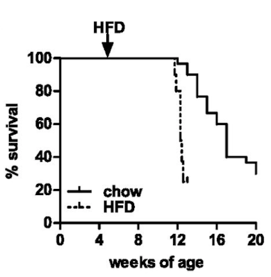
Well, they die. Not unexpected. Here are the survival curves, one group of ATGL knockouts fed chow and another fed an "high fat diet" based on a modified D12492 (mostly extra sucrose with the lard)


If you consider which organ runs (in health) almost exclusively on fatty acid oxidation it will come as no surprise that the mice die of dilated cardiomyopathy secondary to lipid accumulation and mitochondrial failure. Sooner and more rapidly on the high fat diet.

You can get round this problem by engineering the ATGL gene just in to cardiac myocytes. Then the animals live long enough to allow you to study the effects of ATGL deficiency in the absence of a dead myocardium. The whole of the paper, other than Figure S1, uses mice with this protected myocardium (denoted cTg). WT/cTg denotes normal ATGL throughout their body plus extra myocardial ATGL (phenotypically normal) or AKO/cTg without ATGL everywhere other than their myocardium. So ignore the cTg label part, its WT vs AKO re adipocytes throughout the paper

So here's the paradox.


On chow WT mice carry approximately 2g of fat and AKO mice carry 5g of fat, much as the histology suggests and as you might expect. Figure 1 summarises the top left and top right groups of mice:


Things get more interesting when we compare the high fat fed WT mice with the high fat fed AKO mice, thats the bottom left and bottom right. Both groups have increased fat mass but the AKO mice are less obese than the WT mice. Like this


Note that all of the vertical scales are different. But there we have it, knocking out ATGL long term blunts the obesity induced by D12492, somewhat. The effect kicks in slowly but is well established by the end of the study at 22 weeks (solid black squares are AKO)


The paper goes in to some detail about PPAR-γ2 suppression in AKO mice which can be reverse by the diabetes PPAR agonist rosiglitazone.

Which ultimately translates as the adipocytes adapt to being unable to offload triglycerides by suppressing every aspect of lipid uptake and storage that they can.

"... the expression of genes involved in lipogenesis and fat storage such as PPAR-γ2 (−95%), C/EBPα (−30%), and SREBP1c (−78%) were significantly lower in gWAT from HFD-fed AKO/cTg mice than from WT/cTg."

The AKO mouse adipocytes, which cannot off-load lipid, compensate by progressively rejecting lipid ingress.

Does this make the adipocytes insulin resistant? Or the mice insulin resistant?

They didn't look at this at the adipocyte level and the interactions are too complex to guess how adipocytes might respond to physiological or pharmacological exposure to insulin.

What we do know is that the AKO mice fed a high fat diet are still very insulin sensitive at the whole body level despite their adipocytes eventually down regulating all aspects of lipid accumulation. Here's the  intra-peritoneal glucose tolerance test result. All of the following results are high fat diet based.


If you can read Table S1 in the original paper (too faint to reproduce here) you can see that fasting insulin in the AKO mice is 0.1ng/ml vs WT at 1.0ng/ml. Fasting glucose is also low at 164mg/dl in the AKO mice vs 212mg/dl in WT. Sorry for all the Noddy units. HOMA-IR score would be very, very low for AKO mice.

It doesn't matter what size the adipocytes of an AKO mouse are, they are not going to perform basal lipolysis. Under fasting conditions in normal mice FFAs go up as augmented lipolysis frees FFAs and there is little insulin to limit further lipolysis and FFA release. We need elevated FFAs under fasting.

In Table S1 again the WT mice have a fed FFA level of 0.72mM which rises to 0.93mM on a 4h fast, as it should do. In the AKO mice fed FFAs are 0.65mM and drop to 0.45mM on a 4h fast. They drop on fasting, so we can assume that the initial 0.65mM fed value is largely diet derived and so, with no food and no basal lipolysis, FFA levels have to fall.

Which they do.


My premise from Protons is that insulin resistance is an adaptive response to the delivery of FFAs. Under fasting this is ideal. In the presence of elevated glucose and insulin then the elevated FFAs from distended adipocytes cause caloric oversupply to the whole body and insulin resistance has to kick in to adapt. It is an antioxidant defence mechanism to limit ROS generation to physiological levels.

No ATGL -> perennialy low FFAs -> no need to resist insulin -> insulin sensitive

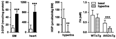
If we look at the hyperinsulinaemic clamp data we can see that both skeletal muscle (SM) and heart in AKO mice are really good at taking up 2-deoxyglucose. The liver, on a diet of 28% sucrose by weight, is also *very* insulin sensitive, with near total suppression of hepatic glucose production (HGP) during the clamp:


Of course the interesting bar chart is the right hand end one. The basal FFA levels are the ones cited above after a 4h fast. Hyperinsulinaemia  with normoglycaemia lowers fasting FFAs a little, but without statistical significance, in WT mice obese from D12492. Doing the same in AKO mice produces a marked fall from low levels to even lower levels, probably somewhere around 0.2mM.

Clearly the excess of plasma free fatty acids, derived from elevated basal lipolysis and which necessitate insulin resistance ie trigger metabolic syndrome, is not present in the AKO mice. Whatever the size of their adipocyte lipid droplets there is no fatty acid release. My guess for the fall in FFAs is that residual post prandial FFAs are being allowed in to muscle and liver cells using CD36 translocated to the cell surface in parallel to GLUT4s in response to the clamp.

Asides before I finish: What does the term "hypophagia" in the title of the paper actually mean? It means that the mice are NOT HUNGRY. They eat ad lib until they are satiated. Because they have down regulated their ability to "sequester" calories in to adipocytes, they sense adequate calories are available earlier so stop eating earlier. They are not "hypophagic", their lack of hunger is manifest as eating less. They're not under-eating. They're eating exactly the correct amount of food to supply their metabolic needs. It doesn't matter that the food appears to be hedonistic, rewarding or addictive (stop sniggering) as it appears to be in the WT mice. Under exactly the same hedonistic/rewarding/addictive food environment (you really must stop sniggering, and so must I) as the WT mice the AKO mice are simply NOT HUNGRY. The brain is such a secondary organ compared to the adipocyte.

Another aside: Where does hepatic insulin resistance in fructose fed mice (like the WT here) come from? Look here


Yes. Fructose, if it gets as far as adipocytes, forces FFA release. This will use ATGL. These FFAs will end up in the liver and have to be repackaged as VLDLs to be returned to the adipocytes. If insulin sensitivity is pathologically high (ie linoleic acid exposure) those FFAs in the liver will be stored there giving fatty liver, NAFLD etc. The AKO mice will obviously catabolise fructose without any problem but will be incapable of fatty liver because they cannot transfer fatty acids out of adipocytes to get to hepatocytes. Hence the incredible ability to suppress hepatic glucose production during the clamp in AKO mice. See HGP in the above figure. Adipocyte AGTL is essential for NAFLD on an high fructose diet.


Okay, I'll shut up now. The role of ATGL in converting linoleic acid induced insulin sensitisation in to whole body insulin resistance and metabolic syndrome is central. This extends to NAFLD and ALD.

Physiology is comprehensible.

Peter

I was going to hit post but two more asides have presented themselves to my brain. Rosiglitazone more than eliminates the down regulation of PPAR-γ2 in AKO mice to give a slightly more obese mouse than the WT high fat fed mice. Does this restore insulin resistance too? Of course not, those bigger adipocytes still can't do lipolysis. The group must know this so they simply didn't run IPGTTs on the rosi-fat mice.

Also re adipocytes under clamp conditions. They looked at behaviour of skeletal muscle cells, heart cells and liver cells under hyperinsulinaemia. But not adipocytes. They know these adipocytes are resistant to insulin, so they didn't check. In a paper on AGTL and adipocyte function. They know this. I love glaring holes from carefully crafted methods sections where you can see the not-investigated leverages.

Oh, and at the time of this study they were clearly looking to find a drug to recreated the benefits of AGTL knockout on weight gain (for high PUFA, high sucrose fed humans). If they had found one I guess they would just have crossed their fingers that it wouldn't trigger dilated cardiomyopathy.

Oh, and another. High PUFA, high sucrose diets clearly do not trigger insulin resistance in the absence of pathologically distended adipocytes forcing elevated basal lipolysis and caloric overload induced high delta psi. The high linoleic acid only generates the 4-HNE to augment insulin resistance and ultimately shut down ETC function when caloric supply and delta psi is excessive. These AKO mice have access to obesogenic levels of PUFA and 21% atmospheric oxygen, yet essentially zero insulin resistance. 

Now I really will stop.

Sunday, March 05, 2023

How can insulin resistance cause weight gain? (5) MCTs

The problems posed to the Protons hypothesis by the failure of fully hydrogenated coconut oil to suppress obesity in rodent models continues to draw me. In the recent past I've gone to Pubmed and searched on "octanoate + ROS" and "coconut oil + ROS".

This paper is pure confirmation bias for saturates resisting insulin signalling via ROS:


This is from 2006 and the software for identifying and quantifying changes in cell appearance was not available (and looks like colour imaging wasn't either!). Now, these are 3T3-L1 "adipocytes" which are the "sort of" fat cells that many people work with in obesity research. 


The white signifies ROS generation. Upper left is control and upper right is with added 1000μM pure octanoate. So a model, using fasting levels of FFA of a composition never seen in vivo. But it generates lots of ROS which can be normalised by adding N-acetyl cysteine. Not only that but it inhibits lipogenesis too:


Bear in mind that you can show almost anything you like by adjusting the conditions during the development of 3T3-L1 adipocyte-like cells, but at face value octanoate is a potential weight loss drug. They didn't look at insulin signalling per se but reduced adipogenesis combined with high ROS generation implies ROS mediated insulin resistance. To me anyway.

The exact opposite is encapsulated in this paper:


Here they used rat brain neurons in culture and used a mix of fatty acids from coconut oil giving in the region of 100μM of mixed, mostly medium chain length fatty acids. So slightly different. We can ignore all of the data relating to Aβ because it's irrelevant and I think that's the wrong paradigm for Alzheimers disease anyway. What I want are the data relating to ROS generation from medium chain triglycerides. This is what they found:


Okay, 24h of incubation with mixed, mostly MCT fatty acids has no affect on ROS. If there is any trend it is downwards. From the Protons perspective reduced ROS lead to preservation of insulin signalling when it should be limited, ie reduced ROS allow excess caloric ingress. 

So look at this. Amongst the many things they measured they looked at the phosphorylation of AKT and it was increased. More insulin signalling will facilitate more fat storage, under fixed conditions:


So what are the differences between the two studies?

In this study these cells are being grown in Neurobasal™ Medium  which comes with the usual 25mM of glucose, enough to support the growth and replication of cells in the complete absence of fatty acids. Some degree of insulin signalling is on-going because it's routine to add supplement B-27 which specifically contains insulin. How much insulin is a trade secret but it is clearly a reasonable amount to ensure effective utilisation of glucose for growth but is unlikely to provide pharmacological levels of insulin exposure which would generate insulin resistance per se. This gives a certain level of phosphorylation of AKT as an effect of that insulin. That's the control cells. Simply adding 100μM of mostly MCTs increases insulin signalling at the pAKT level.

We are talking physiological insulin levels with high glucose and a little coconut oil, compatible with normal conditions after a carbohydrate based meal.

If we go back to the initial octanoate paper we are now talking about the same 25mM of glucose, ie higher than recently fed levels, plus a "fasting" exposure to 1000μM of octanoate. Oh, and 170mM of insulin (that's that 170 times peak post prandial level from previous discussions). This mix is the equivalent of the inability to suppress FFAs under high glucose/insulin, aka loss of metabolic flexibility, aka metabolic syndrome.

Here these adipocytes are awash in a sea of calories and an absolutely supra maximal level of insulin. I think it is reasonable to suggest their electron transport chains are screaming and their need to resist insulin is marked. Using lots of ROS. Almost certainly generated by high delta psi.

What might this tell us?

Under semi-physiological conditions adding even relatively small amounts of mostly medium chain fatty acids increases the phosphorylation of AKT, at a fixed level of insulin. Obviously, to anyone other than myself, that increase in pAKT would be a Good Thing. Insulin sensitising is good, yes? Recall this one:

Small amounts of dietary medium-chain fatty acids protect against insulin resistance during caloric excess in humans

with the effect of eliminating the correct physiological resistance to insulin signalling under forced overfeeding of humans:


Both solid columns are the control state, left hand hatched brown column is the increased insulin resistance of overfeeding fat and the green hatched column on the right is overfeeding very similar fat but with partial replacement by MCT lipids. You can avoid insulin resistance from overfeeding mostly saturated fat using MCT lipids. "Stopping" this insulin resistance is considered a Good Thing.

Except enhancing insulin signalling enhances fat storage.

Summary: The closer you get to physiological conditions the more the suggestion is that medium chain triglycerides facilitate insulin mediated fat storage.

Random thought: You have to wonder if they also stimulate overall growth via facilitated insulin signalling. As in milk fatty acids might be designed, along with milk proteins, to turn baby cows in to grown up cows. Ditto breast fed baby humans.

"How" is a much harder question. Right from near the start of Protons I have accepted at Dr Speijer's idea that very long chain fatty acids, especially saturated VLCFAs go to peroxisomes because mitochondrial oxidation would generate toxic levels of ROS. Later it dawned on me that VLCFA PUFAs probably go to peroxisomes too as they would generate too little ROS for healthy signalling. Now it looks like MCTs carry a different message and are processed differently to longer saturates to provide the specific advantage of maximal growth pre-weaning. Generally adult ROS signalling seems to come from 16 and 18 carbon fatty acids, saturates to resist insulin and MUFA to work in cooperation with insulin. And linoleic acid to muck up the whole system once you get above a certain level of consumption.

There are, if you set your model correctly, parallels between MCTs and linoleic acid of the ROS front. Which leaves me thinking about coconut based cultures with excellent insulin sensitivity but no obesity.

Interesting things, MCTs!

Peter