Monday, January 23, 2023

Delta psi and insulin and ROS

The hypothesis paper

New Control of Mitochondrial Membrane Potential and ROS Formation – A Hypothesis

mentions some factors which might flood mitochondria with Ca2+. Vasopressin gets a mention, again an excellent pressor drug for intractable hypotension, if you don't mind the calcium.

But of course the hormone we all want to ask about is insulin. So you go to Pubmed and search on "insulin calcium ROS"

which brings up this as pretty much the first hit


I haven't read the paper, all I wanted to know was whether insulin behaved as a "stress" hormone as regards Ca2+:

"The insulin-dependent Ca(2+) released from IP3R of skeletal muscle also promotes mitochondrial Ca(2+) uptake."

A lot of cautions yet again. It's neural tissue, which is not a typical insulin sensitive tissue and the paper is old enough that measuring mitochondrial membrane potential was a bit more difficult than ordering a fluorescent dye kit form some generic laboratory supply company.

However it does seem that in something resembling real cells that insulin not only increases delta psi but it also increases ATP levels. Of course we don't know whether the extra ATP comes from glycolysis or increased ox phos from this paper. We do know that it increases.


The insulin concentration used here is 1.0nM which is essentially equivalent to maximal physiological concentration in the aftermath of a meal of modern junk food. Here is the pattern of mitochondrial hyper polarisation at exposure to increasing insulin concentrations. Using 0.75nM is absolutely physiological (if you eat junk food). The glucose used appears to be in the region of 10mM (Ham's F12 medium) and the medium is serum (ie fatty acid) free:


Certain things are clear. There is no dose-response to insulin. Even physiological levels produce a maximal rise in membrane potential. If the pre-insulin membrane polarisation is around 100mV (the technique to assess polarisation couldn't give an absolute value in 2004) then insulin will double this. That seems pretty well certain to generate a membrane polarisation well over the 140mV which will generate copious ROS.

Next thing from here (again)


is that insulin signaling, as assessed by the proportion of Akt which is phosphorylated, is also maximal at exposure to insulin at modestly greater than peak physiological levels (here insulin at 5nM with glucose at ~5mM, some bovine serum and glutamate):


This leaves me with an unanswered question.

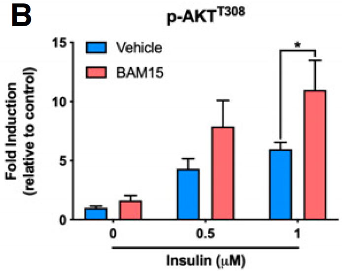
We know that we can increase Akt phosphorylation during massively supra-physiological insulin exposure by simply limiting delta psi with agents such as BAM15 or DNP. This is because we limit the ROS generation which is needed to induce insulin-induced insulin resistance, allowing a little extra pAkt to be formed before ROS exceed a critical threshold. We also know that DNP at in-vivo concentrations does the opposite, it reduces insulin signaling. I won't  re-cite the same old papers. 

What I would like to know is what the oxidation of linoleic acid does to pAkt levels under physiological insulin exposure, compared to palmitic acid. If it is the generation of ROS which limits the rise in pAkt then the lower ROS generation under linoleic acid oxidation should allow more pAkt formation, with enhanced insulin signalling under physiological conditions, which is essential for the development of obesity.

One of my core tenets is that LA limits the normal resistance to insulin signaling mediated by ROS generation. My opinion is the LA causes insulin resistance only once insulin signaling augmentation has produced distended adipocytes which release FFAs in the face of elevated insulin/glucose. In the pre-obese state LA facilitates insulin signaling. Otherwise you wouldn't get fat.

To my knowledge no one has looked at Akt phosphorylation when specific fatty acids are being metabolised under reasonably physiological insulin and glucose concentrations. It would be great to know if LA allowed more pAkt to be formed.

I'd guess that this would be the case.


I have a few more speculations about FFAs, glucose/insulin and ROS which I might leave for another post as this one is getting unwieldy, yet again.

Peter

Saturday, January 21, 2023

Complex IV and control of delta psi

There was a time, quite early in my anaesthesia training, when we used to use a calcium infusion to support blood pressure in anaesthetised horses. You got a bottle of calcium borogluconate marketed for treating milk fever in cattle, hooked it up to a giving set and chose a ball park drip rate by eye. It was bloody effective, easy to use and dirt cheap.

Then we learned a bit more about the role of Ca2+ in cell death and stopped doing it. It's still worth thinking about why it worked.

I have accepted various concepts about the acute control of delta psi and ROS production when metabolic substrate is supplied in excess of metabolic needs. The basic idea is that a replete ATP pool allows delta psi to rise and generate ROS. The earliest ref I've got in support of delta psi and ROS comes from Skulachev in the late 1990s.

High protonic potential actuates a mechanism of production of reactive oxygen species in mitochondria

This is not a physiological model, it just looks at how manipulating delta psi with substrate/inhibitors controls ROS generation. Peak delta psi specified on the graph with their voltage sensitive dye appears to be around 170mV.


The peak values of delta psi and ROS generation are under succinate oxidation and delta psi is modified using either an uncoupler or complex II inhibitor, so, as so often, we are a long way from physiology here but the general principle that ROS generation rises rapidly above a threshold delta psi appears to hold good today. Currently the rise in ROS is thought to occur at around 140mV. Next we can think about the control of ATP synthesis by complex IV, synonymous with cytochrome c oxidase.

This is an interesting review/hypothesis paper from 2001 but I think it too is now quite well accepted:


Peter Mitchell's original concept, to which I have long-term "subscribed", was that electrons passed down the ETC to oxygen, generating a proton gradient, which generates ATP via ATP synthase. If the proton gradient becomes high enough it is no longer possible for electrons to force the extrusion of any more protons (or to be able to flow down the ETC to oxygen) so respiration slows. This appears to be real and to happen at a membrane voltage of 140-200mV. I've extracted the two components of Figure 4 in to separate graphs for a clearer discussion.

Like this:


The red line is the rate of respiration through complex IV as a function of the delta psi generated. As the membrane voltage increases ATP synthase starts turning at around 60mV (the blue line). At just over 100mV ATP synthase activity is maximal and doesn't increase with increasing membrane voltage (in this model). What does increase are those aforementioned ROS generated above 140mV. 

Summary so far: The very high membrane voltages needed to inhibit respiration at complex IV will cause excess ROS generation. This is on the border between physiology and pathology.

There is a second system to control respiration through complex IV. This system monitors the ATP:ADP ratio and limits respiration (and membrane voltage to minimal ROS generating levels) based on rising ATP levels. Like this:


The blue line of ATP synthase activity is unchanged. The green line of respiration though complex IV, as soon as ATP synthase starts to generate ATP, begins to drop and limits respiration though complex IV with a maximum membrane potential at around 120mV, well below that 140mV needed for ROS generation.

So we can limit respiration by inhibiting complex IV using this system at a membrane potential below 140mV with limited ROS generation or we can inhibit it at above 140mV accepting ROS generation using the Mitchell concept. Both are available.

Physiology "chooses" which system to use depending on the circumstance it is presented with. Neither is an "accident". The behaviour of complex IV is determined by it's phosphorylation state. Mostly it's phosphorylated and so behaves like the green line from the second graph and ROS are minimised.

If you strip away the phosphates from complex IV it behaves like the red graph and allows a membrane potential of 140-200mV, with associated ROS generation:

"The allosteric ATP inhibition of cytochrome c oxidase is switched on by cAMP-dependent phosphorylation and switched off by [Ca2+]-induced dephosphorylation of the enzyme (Bender and Kadenbach, 2000)."

That's right: Ca2+ ions dephosphorylate complex IV to allow respiration to proceed to a higher membrane voltage with the acceptance of high ROS generation. The gain appears to be the ability to generate more ATP under "stress" situations and this is primarily under hormonal control. Hormonal control is interesting to look at in another post. But for now:

My bottle of calcium borogluconate was stripping phosphates off of complex IV to allow more ATP production in a myocardium poisoned with an inhalation anaesthetic agent, halothane back in the day. The cost would be increased ROS and it's probably a good idea that we stopped doing it.

Peter

Monday, January 16, 2023

Transformer p245 and onwards: glutathione

In Transformer Nick Lane has an interesting discussion of the roles of glutathione in redox chemistry. He seems to have been spurred to look at this when his research group had a WTF moment with fruit flies. They've not published the work yet so Transformer is the only place to read about it.

They had highly, highly inbred (identical) fruitflies with respect to nuclear genes. They maintained them as identical as possible. Different strains had mitochondria with minor variations in mtDNA, within normal limits, but with differences of "fit" to the (all identical) nuclear genome. Some combinations gave better ETC characteristics than others.

To see if the variations in function were due to ROS flux they treated the flies with n-acetyl-cysteine, a glutathione precursor, producing an excellent scavenger of ROS. You know:


2G-SH  +  H2O2  ->  G-S-S-G  + 2H2O


and all should be well, or at least better.

That didn't work out too well. Males were more or less OK. Most females were "seedy" on NAC. In one strain all of the females, only, died. They checked glutathione levels and the NAC appeared to be doing what it was supposed to do, lots of glutathione.

Lane's idea, which is rather insightful as regards life, is that a body will tolerate an ROS flux within certain limits. To limit excessive ROS formation cells are willing to limit oxidative phosphorylation by deactivating complex I. Compromising ATP production is considered acceptable in order to limit ROS generation to "tolerable" limits.

This is not really surprising. If we think about uncoupling proteins their core function is to dissipate delta psi to a voltage which will not generate many ROS (less than ~140mV), at the cost of decreased ox-phos.

At a guess you might be able to limit/localise the glutathiolation effect to those proteins which are responsible for excess ROS, so glutathione glutathiolates the cysteines within said protein (in this example complex I) in proportion to ROS being generated. Like this


G-SH  +  Prot-SH  +  H2O2  ->  G-S-S-Prot  +  2H2O


Glutathiolation has evolved to alter the function of a protein in such a manner as to decrease ROS generation, even if that includes a decrease in oxidative phosphorylation.

Lane's guess is that female flies, with their high demand for ATP for egg production, couldn't cope with the drop in ox-phos mediated by glutathiolation of complex I.

These flies died in order to limit ROS production.

Have I ever mentioned that ROS are central to, well, everything?

Peter

Supplementary thought: Perhaps we could better phrase it that oral NAC raises glutathione to levels in excess of those which are already ideal, so hyper-glutathiolation causes death by excess ox-phos limitation. This would be particularly problematic for flies with slightly more ROS generation than others. The flies were okay provided ROS and glutathiolation of complex I were at physiological appropriate levels.

Fascinating in view that NAC/glutathione appears to be a pretty Good Drug in general terms. But, as always, over riding evolution has costs.

Sunday, January 08, 2023

Faking it with selenium

 I've looked at this paper in the past, here and here and it is, to say the least, a little dubious in places

High selenium impairs hepatic insulin sensitivity through opposite regulation of ROS

and I really, really want it to be genuine because it supports this hypothesis

High selenium -> high ROS scavenging -> impaired insulin signalling   -> adipose lipolysis

A follow on from this, also plausible, is that

High adipose lipolysis -> excess FFAs to liver -> hepatic ROS from beta oxidation -> hepatic insulin resistance

which would be normal under high FFA oxidation in hepatocytes. I'm willing to accept that reducing ROS in adipocytes might increase ROS in hepatocytes, at a push. But...

These are images taken of hepatocytes from recently euthanased rats fed supplementary selenium for six weeks. This is section A from Figure 3 as a simple copy paste:


What we are comparing is the colour intensity of MitoSOX, a marker of ROS generation,  between control square and the HSe, high selenium square. These two:


Control is essentially black, ie no ROS production and HSe is red, lots of ROS production. Let's cut out the intermediate LSe row and abutt the HSe to the control to make this as obvious as possible:


Now I have another problem. The bright blue colour is an Hoechst stain developed to show DNA, ie it shows the nuclei. I would expect the same intensity of staining with Hoechst stain irrespective of exposure to selenium for 6 weeks or not. The control Hoechst stain is dull in the control while in the HSe image it is bright. Perhaps there has been a problem with the photography?


I can help out by simply increasing the exposure factor for whole of the control row using the colour adjustment in Preview software. This is what it looks like:


Which makes for an interesting comparison between the MitoSOX fields at this (???corrected???) exposure/brightness:


I defy anyone to see any difference between the control MitoSOX intensity and the high selenium exposure MitoSOX intensity. Which makes the rest of the paper, attempting to explain this non-finding, of little value.

So what do I make of this table:


showing a clear dose response to sodium selenite supplementation in both bodyweight and fat weight? For someone who thinks that markedly reducing ROS in adipocytes should cause lipolysis this is exactly what you would expect.

Bear in mind that these rats were gavaged with selenium, it wasn't in the food, there is no "palatability" issue.

I think this is real. Why? Because I want it to be and because of this paper


which found exactly the same effect. This is food intake per week:


The results for weight gain are exactly as expected from the food intake and are given in Table 4, which is huge and turned on its side over two pages. You need a VPN and a line to a server facilitated from Kazakhstan or just take my word for it, food intake predicts final weight, this time.

This is explained away by the authors using ad hoc hypothesis number 12,352 thus:

"A reduced dietary intake was noted throughout the observation period for all treatment groups, which may be considered as a consequence of the unpalatability of the dietary mixture. This resulted in a decreased bodyweight gain in all treatment groups, particularly the females."

I guess, if you were dumb enough, you could call this "reverse Reward". No sniggering!

Combining parts of both papers leaves me deeply embedded in my own confirmation biases, which make sense, at least to me.

ROS are fundamental.

Peter

Friday, January 06, 2023

BAM15 and semaglutide are not insulin sensitising

I'm interested in how an uncoupling agent, which works by limiting ROS generation from the mitochondrial inner membrane, can be described as being insulin sensitising while causing fat loss. This clearly doesn't make sense: anything which increases insulin signalling should increase insulin action. A core action of insulin is the storage of lipid. Fat loss is synonymous with reduced insulin signalling.

This next paper

BAM15‐mediated mitochondrial uncoupling protects against obesity and improves glycemic control

is one I've talked about before. Today I would like to examine Figure 3 in some detail. This work was done using C2C12 myotubes as muscle surrogates and, as far as I can make out from the methods, the DMEM is high glucose, 25mmol/l when fresh, and changed every other day.

The core marker for insulin signalling in the study was the phosphorylation of AKT, shown in graph B of pAKT 10 minutes after exposure to insulin:


I think we can accept that insulin increases the phosphorylation of AKT, blue columns, but there is only a relatively weak dose response in normal C2C12 myotubes. The pink columns show the effect of 16 hours of pre-incubation with BAM15 before exposure to insulin for 10 minutes. I think we can see a reasonable dose response this time with the highest dose of insulin giving the greatest pAKT increase.

Conclusion: Pre-incubation with BAM15 increases pAKT, a good indicator of insulin signalling. Ergo BAM15 is insulin sensitising.

Well, maybe.

Now it's time to re-label graph B. Digging back in to one of Kevin Hall's papers I found that peak insulin in a normal human being after a high carbohydrate meal was in the region of 1000pmol. So we have 0.5micromol insulin as a 500 times peak physiological value and 1.0micromol is 1000 times peak physiological.


Let's be clear. If we use 1000 times peak human physiological insulin we can further increase the action of insulin to phosphorylate AKT by preincubating with the uncoupler BAM15. Clear cut.


Next we should re-visit insulin-induced insulin resistance from back in 2018 and this paper:

Insulin Resistance Induced by Hyperinsulinemia Coincides with a Persistent Alteration at the Insulin Receptor Tyrosine Kinase Domain

and have a look at Figure 2, graph C, which looks like this


I've added the insulin concentrations, this time in nanomoles, which makes it simple to realise we are looking at 5x, 17x or 170x peak physiological insulin exposure.

Core concept: You cannot increase pAKT above that of modestly supramaximal (5x grey squares) by increasing insulin exposure to lethal overdoses, be that 170x physiological here (black squares) or 1000x physiological as in the BAM15 study. Particularly at the 10 minute mark.

Unless you pre-treat BAM15.

My conclusion is that BAM15 increases the level of pAKT induced by massively supramaximal insulin by reducing insulin-induced insulin resistance.  This means that the normal physiological resistance to excessive insulin exposure, ie the refusal to respond further, is blunted. So pAKT goes up.

Again, to clarify: Excessive insulin signalling leads to insulin-induced insulin resistance. BAM15 *reduces* insulin signalling allowing a massive excess of insulin to do a little more phosphorylation of AKT before resistance kicks in. This is a direct consequence of reduced insulin signalling plus supra maximal insulin exposure.

If we were to look at pAKT under conditions of therapeutic uncoupling, it would be decreased. No one in their right mind would do such a study because it would not fit in with the paradigm that insulin sensitisation is a Good Thing. But, if you look hard enough, you can find papers which report this correct finding almost accidentally...

Old stuff again from a blog post last year citing a study from 2015

The Mitochondrial Uncoupler DNP Triggers Brain Cell mTOR Signaling Network Reprogramming and CREB Pathway Upregulation

The study used therapeutic levels of DNP (the mice didn't die!) to uncouple the mitochondria of brain cells exposed to physiological concentrations of insulin (ie produced by eating crapinabag mouse chow):

"The protein levels of AKT, p-AKT (Thr308), ERK, and p-ERK were examined by immunoblotting which showed that the activated (phosphorylated) forms of these kinases (p-AKT and p-ERK 42/44) were reduced in the cerebral cortex at 24 and 72 h after DNP treatment (Fig. 3c–e). Collectively, these results suggest that insulin receptor signaling is suppressed in cerebral cortical cells in response to mild mitochondrial uncoupling."

Aside: I can do exactly the same analysis using metformin. Using a lethal dose of metformin combined with a lethal doses of insulin results in increased peak pAKT.


discussed here giving a therapeutic dose of metformin to a real live human with genetically limited insulin signalling. It reduces their ability to insulin signal still further (and pisses them off big time).


End aside.

BAM15 or DNP (or metformin) all work therapeutically to reduce insulin signalling in vivo. This blunts the insulin signalling needed for insulin-induced insulin resistance in vitro which allows a modest increase in pAKT under massive insulin overdose. Therapeutically this reduced insulin signalling allows lipolysis and fat loss.

Ultimately life has to make sense. Mostly it does.

Oh, and semaglutide with its induction of UCP-1 gene expression is no more insulin sensitising than BAM15. Otherwise it wouldn't give fat loss!

Peter

Tuesday, January 03, 2023

GLP-1 agonists

It was Bill Lagakos who tweeted this paper recently:


The drug itself is of no interest as it will, without doubt, result in Unintended Consequences. But the GLP-1 agonists do seem to work as weight loss/diabetes management drugs, which is strange. The paper per se describes clinical work and looks to be written by clinicians so is unlikely to provide any sort of insight as to mechanism(s) of action.

It is worth thinking about how a drug which increases insulin synthesis, insulin reserves and insulin secretion in response to glucose might actually cause weight loss. Really, it shouldn't... Everything is against this. But it does.

it appears that the GLP-1 agonists activate the formation and preservation of new adipocytes, much as the glitazones do:

"These findings, combined with our results, strengthen the notion that GLP-1 or liraglutide regulate adipogenesis via suppression of apoptosis and stimulation of proliferation."

So the GLP-1 agonists stimulate adipose hyperplasia meaning you develop lots of small adipocytes which are much better able to act as a sump for calories. There appears to be a shift of fatty acids out of pre-existing hypertrophied adipocytes, and out of ectopic fat deposition sites too, and in to these spanking new baby adipocytes.

Why don't you get hungry and fat, as you do with the glitazone drugs?

You should do.

This paper used ob/ob mice (so caution) 

GLP-1/GLP-1R Signaling in Regulation of Adipocyte Differentiation and Lipogenesis

and confirms the presence of those small adipocytes in white adipose tissue under GLP-1 agonists, a lack of visceral fat hypetrophy and, interestingly, a down-regulation of the gene for the fatty acid synthase enzyme, crucial for de novo lipogenesis. Not only are the mice used ob/ob but the "high fat" diet is unspecified, even following all three refs they cite as describing it. So even more caution about these people. But in both 3T3-L1 "adipocytes" and in real ob/ob adipocytes, fatty acid synthase gene expression is down regulated. Their suspicion is that suppressed de novo lipogenesis limits adipocyte hypertrophy. They have no insight as to why.

And what regulates fatty acid synthase gene expression? Did I hear insulin from the back there? I hope so.

Let's summarise:

GLP-1 agonists induce the production of large numbers of small, highly insulin sensitive adipocytes which don't become distended. They don't distend (probably) because fatty acid synthase gene expression is down-regulated. I consider this to be a flag for suppressed insulin signalling. Suppressed insulin signalling leads to a lean phenotype. So an insulin sensitising agent reduces insulin controlled gene expression to avoid obesity. WTF?


After this preamble let's get to an abstract which was the first hit of my PubMed search based on GLP-1, adipocyte and ROS:

I don't have the full text but the abstract alone is quite informative. Here are the relevant statements:

"Semaglutide enhanced multiloculation and uncoupled protein 1 (UCP1) labeling in obese mice [adipocytes] ..."

and

"Besides, semaglutide activated adipocyte browning, improving UCP1, mitochondrial biogenesis, and thermogenic marker expressions help weight loss."

I think it is reasonable to accept that GLP-1 agonists activate uncoupling. We have know for a very long time, since the days of di-nitrophenol and more recently BAM15, that uncoupling reverses metabolic syndrome.

For the time being I will just leave it that GLP-1 agonists decrease insulin's action secondary to uncoupling. In this they are similar to the uncouplers DNP and BAM15 and also to metformin which blocks insulin's action, not by uncoupling but by blockade of the glycerophosphate shuttle. All work via decreased ROS generation.

Reduction of insulin signalling is intrinsic to weight loss.

Quite how decreasing insulin signalling via uncoupling can be made to show an increased insulin responsiveness (right down to the level of Akt phosphorylation) is an interesting question. I got part way through discussing this next using a paper on BAM15 but it made the current post so long and unwieldy that it needs a post of its own.

Peter

Throw away comment (I have not been through these papers!). Many people are reported to be using a GLP-1 agonist for weight control. It's probably working by activating/generating UCP-1, which is cool. At the same time it is generating lots and lots of new baby adipocytes, which stay small through uncoupling.

Currently the risk of promoting breast cancer by GLP-1 agonists is taken sufficiently seriously to have promoted a systematic review and meta-analysis to bury said risk:

Do GLP-1 Receptor Agonists Increase the Risk of Breast Cancer? A Systematic Review and Meta-analysis

Sadly metabolic reality couldn't give an F about systematic review and meta-analysis:

Glucagon-like peptide-1 receptor activation by liraglutide promotes breast cancer through NOX4/ROS/VEGF pathway

and eventually these drugs will be withdrawn. So what will happen to folks with years of adipocyte hyperplasia, where individual cell hypertrophy has been suppressed? Their very numerous small adipocytes will, without their uncoupling drug, become enormous. People will become hungry as they develop hypertophy of all of those lovely tiny insulin sensitive adipocytes. Oops. It's gonnabe bad, but that's years down the road.

Sunday, January 01, 2023

How incomplete was LUCA?

LUCA is the Last Universal Common Ancestor and she had a number of quite odd characteristics which provide some insight in to what conditions might have been like at the dawn of life on Earth.

I hope we have all watched Lex Fridman chatting with Nick Lane:


Somewhere around 8m 50s they tackle the subject of why the existence of both bacteria and archaea imply life only originated once on earth. Towards 15m they discuss the differences between the two life forms and the very high probability that they both arose within the same localised vent system.

What I felt was missing from the discussion was the incomplete nature of LUCA. It's implied but not explicit. Maybe that's just me and my own biases but the fact that LUCA was incomplete and differentiated in to bacteria and archaea as alternative routes to completion is very central to how life evolved and has something to say about the conditions at the time.

The core reaction at the origin of life is

CO + 2H+ + 2e-   ->   CO + H₂O

The electrons come from reduced ferredoxin, which is a fossilised remnant of the FeS structure of the hydrothermal vent system, where the pH differential across FeS minerals, between vent and oceanic fluids, provides electrons (and hydrogen) with the power to reduce CO. I went over it in horrible detail back here.


and shows exactly the core reaction at the origin of life, it runs from bottom to top.


We also know from Huber and Wächtershäuser's work

Activated acetic acid by carbon fixation on (Fe,Ni)S under primordial conditions

that supplying a methyl source (they used CH3-SH) and an iron/nickel/sulphur catalyst, that activated acetate was easily formed from exogenous CO (but not from CO, you need the reduced ferredoxin for that step, as above). Thus:

CO + CH3-SH + HO -> CH3.COO-SH   (activated acetate is energetically approximately equivalent to ATP)

In origin of life scenarios the initial supply of methyl groups is assumed to be of geochemical origin in the vents. To leave the vents you need a) a source of reduced ferredoxin to generate the CO from CO and b) a source of methyl groups to replace the geochemical CH3-SH. Oh, and some hydrogen but thats another story.

Archaea and bacteria have solved the problems of ferredoxin supply and methyl supply in completely different ways. Neither could have left the vents without solving these problems. Which means that LUCA was dependent on geochemical CH3-SH and the two derivatives of LUCA escaped the vents by different techniques for its replacement. But ribosome function, RNA replication, DNA synthesis (but not duplication) are so similar that it is almost certain that both escapes occurred from the same overall vent population. These then spread out to other vents and took over all of the early Earth. If other vent systems were in the process of developing "life" then that life lost out to archaea and bacteria and their shared early features. LUCA is thought to have had somewhere around 30 core enzymes.

When you pick up papers on the phylogenetics of various proteins which can be traced back to LUCA by non-Lane-ophiles you have to mentally appreciate quite how limited LUCA was and how early in evolution LUCA differentiated in to archaea and bacteria. This happened while still trapped an oceanic alkaline hydrothermal vent. That's how far back LUCA lived, fixed in one location.

So it's sort of odd that oxygen centred enzymes such as catalase, superoxide dismutase (iron based, FeSOD) and the globin oxygen binding protein ancestor (precursor of haemoglobin et al) were all present in LUCA. In an anoxic ocean.

So when I look at ROS signalling I'm thinking of the nature of LUCA, ie a very, very early, pre free living organism.

Peter

Aside: why two escapes? Crude fatty acid membranes and early isoprenoid membranes were both fairly permeable to both protons (from the oceanic side) and OH- ions (from the vent fluid side). This was essential to allow continuous ingress of protons to drive a H+/Na+ antiporter. Fully impermeable membranes would not allow neutralisation of those protons by OH- ions and metabolism would collapse.

Somewhere (I cannot recall where) I read that mixing fatty acid membranes with isoprenoid membranes produces a membrane which is fully impermeable to both protons and OH- ions, shutting down the ability to neutralise those ingressing protons on which LUCA based her metabolism. If one LUCA population invented fatty acid membranes in one geographical area of a vent and another LUCA population, geographically distant but still within the same vent, chose isoprenpoids then the area of mixing of the populations would become an impossible situation for a metabolism based on the necessity of proton and OH- permeability. Which would keep the populations separate, within the same vent, to evolve differing tools to allow escape. End aside.