Monday, May 16, 2022

Deuterium protected linoleic acid

This is a fascinating paper which has distracted me from my thought train on uncoupling, mitochondrial membrane potential and ROS generation because it has aspects involving all three while not being particularly intuitive as to what is going on. I picked it up via comments from Tucker and Raphi on twitter.

Deuterium-reinforced polyunsaturated fatty acids protect agains atherosclerosis by lowering lipid peroxidation and hypercholesterolemia

First aside: Mouse model. This current model, the APOE*3-Leiden mouse, is a model. It's not as totally useless as an APOE total knockout or an LDL receptor knockout model but it's still nothing like a real mouse or like a real human. Mouse lipoprotein management is not like human lipoprotein management. They do not have a cholesterol ester transfer protein. The Leiden mouse has this human gene engineered in. It also has a selective APOE*3 knockout to give a mild elevation of LDL. The end result is a model which has numbers on a lipid panel which look a bit more like a human with metabolic syndrome than the average extreme knockout mouse model.

But it's not a human with metabolic syndrome. It's an APOE*3-Leiden mouse and if you found a cure for its "atherosclerosis" you would have a great tool for helping APOE*3-Leiden mice. Would it translate to helping humans with metabolic syndrome? Hahahahahahahahah bonk. End aside.

Second aside: Does LDL cause atherosclerosis? Hahahahahahahah, bonk. To anyone with any sense atherosclerosis is a response to injury where IGF-1 delivered by platelets attaching to the injured endothelium causes media hypertrophy to reinforce the site of injury. This is accelerated if systemic hyperinsulinaemia also acts as an agonist on those IGF-1 receptors. It can almost certainly be enhanced by delivering lipid peroxides such as 4-HNE, 13-HODE and 9-HODE, though their effects are very, very complex. I'm perfectly willing to believe that any genetic engineered tweak in to a mouse which increases the persistence of linoleic acid containing lipoproteins in the plasma allows time for that LA to spontaneously oxidise and accelerate what looks a bit like atherosclerosis, in the model. Just my view. End second aside.


OK. On to the paper:

The APOE*3-Leiden mice were reared on non specific chow. At 12 weeks of age (Time -4) they were put on to something derived from the AIN-93M diet. All lipids were all supplied as methyl esters of fatty acids, not triglycerides. It contained 1.2% of calories as LA and 9% of calories as sucrose. The intervention group had exactly half of the 1.2% of LA calories supplied in the form of deuterium stabilised, ROS peroxidation resistant D2-linoleic acid.

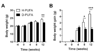
They were fed these diets for four weeks. At that point (Time zero) 0.15% by weight of reagent grade cholesterol was added to both diets (otherwise the model doesn't work to get the essential-for-funding lipid lesions, no sniggering at the back. It's a model). This "western" diet, which only differed from the run-in diet by the added 0.15% reagent grade cholesterol, caused/allowed some weight gain over the following 12 weeks but less in the D2-LA supplemented group than the normal LA group:


No weight gain on either of the run-in diets, followed by lots of gain in the "normal" LA diet but not the D2-LA diet once the cholesterol was added.

Why?

Why should adding 0.15% of cholesterol produce such diverging weight gains?

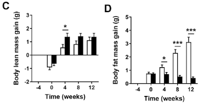
Even more exciting is if you look at lean mass vs adipose mass:


Adding just 0.15% cholesterol produced a marked fat mass gain in the normal LA mice and a trend downward in fat mass for the ROS protected D2-LA mice.

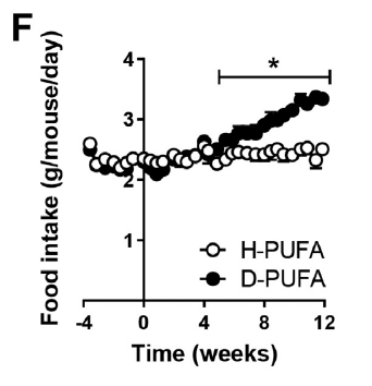
On top of that the D2-LA mice started eating extra during the period of fat loss. A lot extra:


Soooooooo. What is going on?

Well, the first thing to realise is that during the four week run-in period after the replacement of chow by the AIN-93M derived diets there were already changes which didn't show in total body weights (graphs A and B). The mice, with or without deuterium stabilised LA, all lost muscle mass and all gained fat mass during those weeks, just under a gram of each. So, even without the reagent grade cholesterol, changes were already on going from a "normal" mouse phenotype on chow towards a "skinny-fat" phenotype on an AIN-93M-like diet. That's clear in graphs C and D. Possibly from the sucrose but there's no way of telling that from the paper.

The changes were on-going before the diets were "westernised" by the addition of 0.15% of reagent grade cholesterol. I suspect that the addition of the cholesterol is a red herring.

Let's go on to look at food intakes.

The mice on the deuterated LA eventually began eating more than those on the standard LA. This became statistically significant at about week five.


Any mouse which is eating extra and losing adipose tissue is either showing malabsorption or uncoupling.

I'll buy the uncoupling, but then I would.

Why the delay to the onset of starting to eat extra? Is there a delay in uncoupling onset? Not necessarily. A normal mouse uses a significant percentage of its caloric intake to generate heat in its brown adipose tissue. There is no need to increase food intake while ever the purported uncoupling from deuterated D2-LA is generating less heat than is needed to maintain body temperature. As heat production increases over time it begins to exceed this essential minimum and so comes to represent a "calories-out" in excess of what is merely needed to keep warm. At this point an increase in food intake becomes necessary to balance the heat lost by supra-physiological uncoupling.

If this is correct, and that's a big if, there is clearly an on-going progressive increase in uncoupling with time. The logical explanation is that there is a progressive increase of deuterated D2-LA in tissues and/or being used for beta oxidation.

How might deuterated, ROS resistant LA, facilitate uncoupling?

I don't know, so it's time for some routine wild speculation. If we could answer that one question the whole scenario becomes straight forward. Sadly it is not at all obvious why D2-LA should facilitate uncoupling. Here's my current best shot. If I think of something more plausible I'll post again:

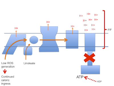
Let's assume that linoleic acid, whether deuterated or native, allows excess calories in to a cell. This is the doodle from a few posts ago:


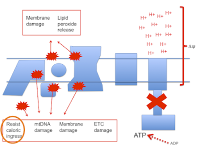
which then leads to this doodle:


and this doodle:


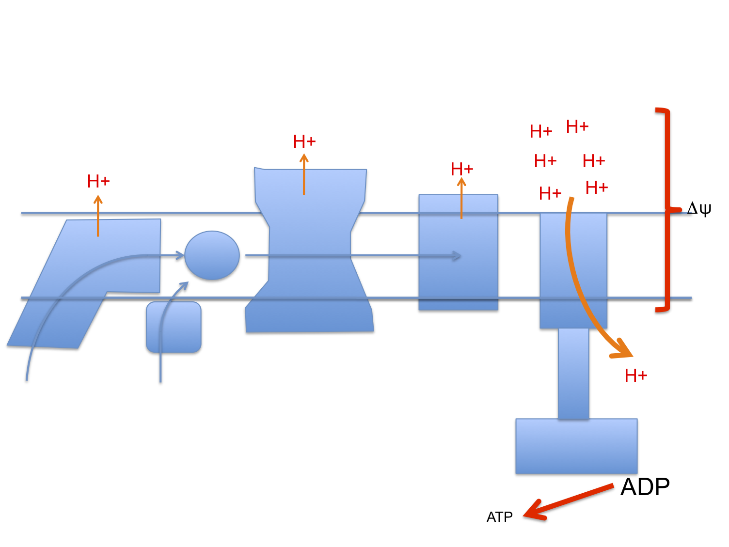
This begs the question as to how much damage (signalling?) is done by the stray electrons, how much by superoxide, by hydrogen peroxide or how much is actually mediated by the lipid peroxides generated from linoleic acid per se. Which is the most important mediator?

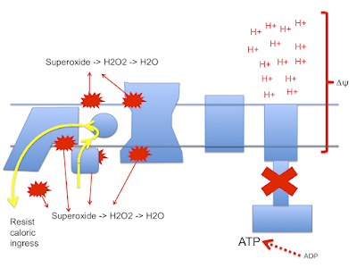
Let me suggest that D2-LA allows the excessive delta psi, which facilitates both reverse electron transport and pathological ROS generation. As in the previous post the high delta psi eventually allows D2-LA to drive RET at the cost of, via high delta psi, allowing electrons to be lost from the ETC to oxygen at abnormal sites, forming superoxide. Under D2-LA this superoxide has very limited ability to contact oxidisable native linoleic acid.

So now we look more like this with ;


There is a surfeit of ATP, high delta psi and availability of either LA and/or D2-LA to facilitate uncoupling combined with minimal damage to the ETC. You do have to have a source of 4-HNE or a related "damage marker" to facilitate uncoupling but it doesn't need much.

I can't see any more straightforward technique for D2-LA to uncouple. If there is one and it's clear how it works, that would be great. Currently this is the best I can do.

Summary: D2-LA allows uncoupling. That explains everything, but the mechanism for the uncoupling is obscure and I'm guessing.

NB I was also trying to explain to myself why the control group got fat. I don't think they did. A total weight gain of 5g over 12 weeks to give a final weight of 25g sounds like a normal mouse to me. It's the slim mice eating extra food to maintain that slim bodyweight that are abnormal.

Ultimately the paper poses the question: What determines whether a cell deals with excess calories by sequestration to storage vs uncoupling. Obviously this is insulin signalling. But is it an oxidation product of linoleic acid which controls insulin signalling? Are we simply looking at a situation of absolutely suppressed insulin signalling, due to D2-LA being "too" stable?

Peter

Final addendum/aside. There is a claim on 'tinternet, un referenced, that low dose oral deuterium oxide in mammals causes weight loss, rather than the death which is the normal result of high dose exposure. Could D2O trigger uncoupling irrespective of LA type and the catabolism of D2-LA be a simple source of deuterated water by oxidation of the D2-LA? I doubt this but the idea is still a potential explanation. I have hunted support/refutation for this without success.

Sunday, May 08, 2022

Protons (70) Uncoupling does suppress insulin signalling

The premise of the last post is that mild mitochondrial uncoupling is protective against fatty liver because it disables insulin signalling and generates heat.

There is a considerable literature looking at "energy stress" in cells induced by marked uncoupling, usually using dinitrophenol (DNP) to profoundly reduce ATP generation from oxidative phosphorylation. The DNP concentration in cell culture to achieve this would usually be 1mM. Oral dosing can transiently achieve plasma levels in mice of around 0.5mM and is probably higher in the liver because it receives the portal blood flow from the site of absorption in the gut, so maybe this has some application to real life. Maybe. These studies are peripherally interesting as dropping ATP this aggressively triggers AMPK activation which translocates GLUT4 to the cell surface to maintain cell viability using ATP from glycolysis. The translocation is independent of any markers of insulin signalling.

An example from 1988

Evidence for two independent pathways of insulin-receptor internalization in hepatocytes and hepatoma cells


The basic premise is that ATP depletion is the activator of AMPK mediated translocation of GLT4s to the plasma membrane. However AMPK is also activated by both fasting and ketogenic diets, neither of which produces an acute ATP deficit. Years ago I suggested that a major activator of AMPK is acute loss of insulin exposure and/or its signalling, independent of ATP status.

So DNP at 1mM (1000μM) in cell culture does indeed deplete ATP and AMPK does indeed translocate GLUT4 under these circumstances. Is the mechanism of action the acute suppression of insulin signalling (secondary to the loss of mitochondrial membrane potential) rather than, or in addition to, the fall in ATP generation per se?

Happily it is quite easy to measure insulin signalling nowadays. It's also possible to use either live mice taking non-lethal doses of DNP or cell culture using therapeutic concentrations of DNP. 

This next paper used DNP in live mice at non lethal dose rates and in cortical neuronal cell culture at 10-40μM concentration as opposed to 1000μM.

The Mitochondrial Uncoupler DNP Triggers Brain Cell mTOR Signaling Network Reprogramming and CREB Pathway Upregulation

Bottom line: Mild uncoupling using a therapeutic concentration of DNP suppresses insulin signalling. In this case they are looking at whole cerebral cortex in their live (until euthanasia for brain removal) mouse model or cortical neuronal cell culture.

"The protein levels of AKT, p-AKT (Thr308), ERK, and p-ERK were examined by immunoblotting which showed that the activated (phosphorylated) forms of these kinases (p-AKT and p-ERK 42/44) were reduced in the cerebral cortex at 24 and 72 h after DNP treatment (Fig. 3c–e). Collectively, these results suggest that insulin receptor signaling is suppressed in cerebral cortical cells in response to mild mitochondrial uncoupling."

That seriously confirms my biases.

I'm perfectly willing to extrapolate from mouse neuronal cells to mouse hepatocytes because this is a basic principle of how I view energy physiology working. I might be wrong, or not.

Low dose DNP and BAM15 will both treat metabolic syndrome in humans.

Conceptually what is happening in metabolic syndrome at the most basic level is that linoleic acid is allowing too many calories in to a cell and this leads to both storage and pathological ROS generation to side-step and/or limit the process. It is a coping mechanism for the failure of LA to signal physiological insulin resistance cf that provided by palmitate.

Uncoupling suppresses insulin signalling. If there are stored calories within the cell they are then made accessible. These calories are used for ox-phos to make up the deficit caused by the uncoupling. At normal weight (ie without excess insulin signalling due to diet) the suppression of insulin signalling will be accommodated by AMPK activation.

Stuff makes sense.

Peter

Saturday, May 07, 2022

Protons (69) BAM15

Completely at random Tucker emailed me this paper using the classical model of Bl/6 mice fed an unspecified 60% fat diet to become obese. He thought I might find it interesting from the Protons perspective. He is correct.

Mitochondrial uncoupling attenuates sarcopenic obesity by enhancing skeletal muscle mitophagy and quality control

The background to the mitochondrial uncoupler BAM15 is in this paper

BAM15 has the potential to be a blockbuster so I think we should be very, very cautious in how we view these reports of marvellous efficacy. They're probably correct, but caution.

The papers above fit nicely in to the venerable work I've recently been reading relating to DNP, which I'll come back to in a post or two.

Today I just wanted to point out a glaring problem within the BAM15 papers for anyone viewing the work from the Protons perspective.

BAM15 reverses the insulin resistance of diet induced mouse obesity models. As in the last ref:

"Collectively, these data demonstrate that pharmacologic mitochondrial uncoupling with BAM15 has powerful anti-obesity and insulin sensitizing effects without compromising lean mass or affecting food intake." My italics.

I maintain that insulin sensitising makes you fat and that uncoupling should make you thin by suppressing insulin signalling, assisted by a "calories out" component of heat generation.

Let's look at the explanation of how we square this circle.

We need Figure 1 plus the methods section. An oral gavage of BAM15 at 10mg/kg achieves a therapeutic concentration and shows an elimination half life of around 1.7 hours in mice.


 Plasma concentration appears to be sub-therapeutic by four hours max, probably drops too low by two hours. The rest of Figure 1 shows us that BAM15 is essentially liver specific. To demonstrate that the uncoupling is liver specific you have to give that higher dose of BAM15 (50mg/kg) and measure tissue specific oxygen consumption at peak effect,  ie one hour post gavage.


We can also see that by four hours after a therapeutic gavage that liver concentration is also sub therapeutic following a 50mg/kg oral dose:


Then we have the methods section where we can find that the euglycaemic hyperinsulinaemic clamp was performed, as you might expect, after a fast of five hours. The drug is in the food.

By five hours of drug withdrawal the clamp is actually looking at the behaviour of the phenotype induced by the drug, not at the mechanism of action of the drug itself which was used to achieved that phenotype.

If we consider NAFLD as the pathological storage of lipid in the liver secondary to "mopping up" of FFAs released from overly distended adipocytes (with overactive basal lipolysis) then we are in a position to see why suppressing insulin signalling in hepatocytes releases fatty acids from hepatic triglycerides (aka fatty liver). There is no need for export of those released fatty acids in the form of VLDLs because each uncoupled hepatocyte is already in caloric deficit secondary to that uncoupling, which is what is suppressing insulin signalling while simultaneously providing a "calories out" route as heat.

Resisting insulin -> diminishing fat storage.

Continuous disposal of FFAs being stored in the liver from over distended adipocytes, without recycling those FFAs back to adipocytes as VLDLs (aka high fasting triglycerides) produces a slim, insulin sensitive phenotype. Because adipocytes become small, with associated low basal lipolysis.

Just one feature of BAM15, uncoupling, produces both specific hepatic insulin resistance (reducing hepatic triglyceride storage) combined with unstoppable drug-induced hepatic lipid oxidation as a "calories out" route.

Both from that single action of uncoupling. Neither effect is present once the drug wears off.

Insulin sensitivity/resistance being "good" or "bad" is absolutely context specific.

There is no glimmer that any of the above cited papers have an insight as to this being the mechanism of action of BAM15.

But that's how it works.

Support comes from the DNP papers.

Peter

Wednesday, May 04, 2022

Protons (68) Pathological ROS (1)

This is another non-referenced, thinking out loud post which is the precursor to more normal technical posts. Here we go.

The whole underpinning of the Protons concept is that inadequate generation of ROS secondary to the presence of double bonds in fatty acid fuels causes pathological insulin sensitivity.

Too few ROS.

This post is about how generating too few ROS in mitochondria generates excess ROS in those said mitochondria and what physiology does about this.

Here's the stripped down doodle of the electron transport chain I'm going to use


I've left out complex II, cytochrome C etc to keep it very simple. It's not complete, it's a minimal mental "model". You have been warned.

Next is the normal electron flow from NADH and FADH2 to oxygen:


Electrons passing through complexes I, III and IV pump protons out of the mitochondria to produce an electrical and pH difference between inside and out, the mitochondrial membrane potential, delta psi


and of course delta psi is used to generate ATP by ATP synthase, much as a rotating water mill uses hydrostatic pressure to generate usable energy.


One crucial necessity to allow ATP synthase to function is a supply of ADP. If all of a cell's supply of phosphorylated adenine is in the form of ATP there is minimal ADP as substrate for ATP-synthase to act on, so delta psi will become larger as pumped protons accumulate on the outside of the mitochondrial membrane.


If ATP predominates the cell is replete and the correct response is to limit further caloric ingress. As ETFdh tries to transfer electrons on to the CoQ couple and subsequently to complexes III and IV it becomes progressively harder to pump protons against a rising delta psi. At some point it becomes easier to divert electrons from ETFdh through complex I as reverse electron transport (RET) which generates a very specific, localised ROS signal which is designed to limit insulin signalling and be quenched using superoxide dismutase without doing damage. Saturated fats produce this signal very well because their lack of double bonds maximises the input of FADH2 to facilitate RET:


In this mental model palmitate limits caloric ingress, stops excessive proton pumping and maintains delta psi within physiological limits.

Next we have the situation under linoleic acid oxidation. Here there is a reduced input of FADH2 from ETFdh so it is more difficult to generate the RET needed to limit caloric ingress when ATP is replete and ATP-synthase is no longer consuming delta psi. Protons continue to be pumped and delta psi rises to supra-physiological levels


As delta psi rises the ability to generate RET through complex I also rises until eventually even the relatively low FADH2 input from linoleate can produce RET. However this high delta psi will also allow the generation of ROS at multiple sites in the ETC in addition to that at complex I. Complex IV seems to be a minimal site for this but complex III will produce ROS from sites facing both the mitochondrial matrix and the cytoplasm while complex I appears to only generate on the matrix side, probably from multiple sites under very high delta psi. ETDdh (and mtG-3-Pdh and complex II) can also generate ROS under high delta psi conditions:


This is both good and bad.

Good because it finally hits the signalling pathways needed to limit caloric ingress. Bad because it hits lots of other components of the cell structure in addition.


Summary: consuming linoleic acid will cause your mitochondria to explode.

Except that's preposterous, they don't.

The simplest protective measure is the diversion of calories within the cell in to storage. Those calories are only present in the cell secondary to excess insulin signalling. Because diversion to storage is a classical function of insulin signalling, this will drive obesity whilst also providing some protection from pathological ROS generation.

Additional protection comes from uncoupling.

The core mechanism for the generation of excessive ROS under unmitigated LA oxidation is high delta psi.

Uncoupling lowers delta psi. Doing this is all that is necessary to protect against pathological ROS generation.

But wait.

If linoleic acid allows excess caloric ingress due to a deficit in physiological ROS generation, surely non-specific suppression of ROS generation should increase caloric ingress, increase delta psi to overcome the degree of uncoupling present and re establish pathological ROS generation? Alternatively might uncoupling go on to allow even more excess calorie storage?

Neither happens.

I'll run through some of the papers I've been looking at over the last few months and post about them next.

Peter