Saturday, December 10, 2022

ROS and cell to cell signalling in plants

The people who wrote this review are plant biologists. Nothing wrong with that and they've been doing some fairly deep thinking about reactive oxygen species signalling.

ROS signaling: the new wave?

In this paper I'd like to just run through the images in Fig 1 from Box 1 with some sort of commentary.

This is an ancestral organism. It could represent LUCA, which has implications about what LUCA might have needed to do to deal with ROS. Under conditions of rising oxygen tension (or even without rising oxygen tension) there is the need for basic ROS scavenging simply to survive. Any organism which couldn't do this has not left descendants:


If you want to maximise your chances of survival then the co-opting of some sort of protein as an ROS receptor provides an advantage. A better controlled response to suppress ROS to acceptable levels becomes possible without wasting  resources on excessive scavenging systems. The little horse-shoes are the receptors:


This review is from 2015, at which time ROS generation by bacteria was thought to be limited to "accidental" release during photosynthesis by cyanobacteria. Obviously this is no longer the case but anyway, we can think of the blue circle as representing an organism or cell as part of the same organism capable of generating and releasing significant ROS, which our golden organism might not like and might dedicate another sensing/quenching system to:


If the sensing system turns out to be carrying useful information about the neighbouring organism/cell our golden cell might consider it worth the NADPH to send a signal back. Maybe a "come on" signal, maybe a "bugger off" signal. But ROS based too. Here we are talking eukaryotes as the ROS generator (in this review) is considered to be one of the NADPH oxidases (NOX), the small red bauble, which does not appear to be coded for in prokaryote genomes:


Assuming that cell-cell signalling proves helpful, the system can go on to develop as many responses as is useful to survival:


Back to plant biology. This next image summarises what the group thinks happens in groups of plant cells, this time taken from Figure 1 of the text proper:


And because they are plant biologists, and because plants sit still (on a human timescale) when you kick them, here is what happens when you "hurt" or injure an Arabidopsis plant, illustrated using an fluorescence probe. There is a luciferase gene spliced in to the ROS generating machinery. Note the timescales:


So plants appear to signal within the organism, on a timescale which is appropriate to their needs, using ROS and ROS receptors. I've been curious about how an oak tree root hair might signal to the top of the canopy. Even for a plant speed of communication needing several days might be too slow.

Is it possible to convert a localised ROS signal to an organism-wide information network signal?

Maybe.

Peter

Thursday, December 08, 2022

I'm back

Hi all. Well, I'm back.

Over the years I've taken a few breaks from blogging. Eventually something takes my fancy and I restart. There is usually a stack of way too many backlogged comments to reply to and I just go through deleting the frank spam and approving all of the rest.

Which is highly unsatisfactory but is just how it goes. My apologies.

I hit a total brick wall on the roll of medium chain triglycerides in insulin signalling vs resistance. I've not resolved this and still have coconut oil and butter as subjects for long term consideration. Hence the couple of posts over the last few days.

I'm also interested in the absolute primacy of ROS as the underlying signalling system in and between all cells, prokaryotes included. They underly insulin signalling the way machine code (is that still a thing nowadays?) underly Cobol or Fortran.

And if LUCA was, of necessity, anaerobic, where did she get her oxygen from to use to make ROS as signalling molecules?

I have a post part written starting on this front and will tidy it up and post as I get the chance.

Am I well?

I'm still bouldering once a week, just about at V4a on good day. Had a go at roped climbing earlier this week. Enjoying the start of winter here although last summer was marvellous in the UK. Looking forward to the Solstice in just over a week. Run at MAFF heart rate occasionally but not nearly as much as I'd like too.

Still unvaccinated and no idea if I've had the 'vid since I failed to contract it from my wife back in Feb/March 2020. Had a proper cold two weeks ago but it never crossed my mind to do any sort of test. It was a cold, with a good sore throat. Kids are back at school etc so... Shrug. For odd practical reasons the day of its initiation was associated with a one day break from carnivore at a posh do in London. I blame the vegetables!

Life is good.

Peter

Wednesday, November 30, 2022

Sugar on your coconut oil?

I mentioned this paper in the last post:

Soybean Oil Is More Obesogenic and Diabetogenic than Coconut Oil and Fructose in Mouse: Potential Role for the Liver

In addition to looking at soybean oil they also looked at fructose. This is what eating 25% of your calories as fructose in your pelleted food does to your liver. Again red indicates lipid deposition in the liver.

First a reiteration of 5001 vivarium chow:


Then 25% fructose on the high coconut background:


and on  the 10% soybean oil background


So I think we can say that eating 25% of your calories as fructose might be excellent if you are a Bl/6 mouse who wants a fatty liver.

But what does fructose do to your weight/fatness?

That depends. If you are on coconut oil (+2% calories as linoleic acid) it increases your weight towards the weight of the soybean oil group.

If you are on soybean oil then including this much fructose decreases your weight to that of the coconut plus fructose group.

How come?

Sadly feeding mice on 25% of their calories as fructose generates rectal prolapses in 30-40% of those mice. And rectal prolapse is, in all probability, just a surrogate for chronic gut rot.

I would be very, very cautious about interpreting any of the data from mice with this degree of gut dysfunction. Which does nothing to allow me to decide whether the sucrose present in high "fat" diets such as the Surwit coconut/sucrose diet or any of several butter oil/sucrose diets is significant.

Which would be nice to know.

So let's look at a paper by Surwit's group:

Reversal of Diet-Induced Obesity and Diabetes in C57BL/6J Mice

All you have to do to reverse both obesity and diabetes in Bl/6 mice is remove the fat from the diet. 

Here are the compositions. The low fat diet always reversed the problems from high fat in a few months. For Bl/6 and A/J (less obesity prone) mice:


If you glance down through the ingredients things look very similar except for the fat and carbohydrate composition. Right down to the inclusion of exactly 18.3g/kg of soybean oil to avoid essential fatty acid deficiency in each diet.

Aside: Of course most of the components are the same by weight. But not by calories. The high fat diet dilutes everything with saturated fat calories, by about 25%. This includes the linoleic acid from the 18.3g/kg of soybean oil. This is a very low (~roughly 1.6% kcal) LA diet. End aside.

Oh, and the sucrose. When the weights per kilogram of most components of the diets are identical to a decimal place, suddenly 10% of calories as sucrose arrives in the high fat diet. Why? Surwit discusses its inclusion and correctly points out that 10% sucrose in low fat diets does not cause obesity or diabetes in Bl/6 mice. But he does not address why the sucrose was "needed" in the high fat diet.

I leave you to speculate as to whether the sucrose is essential to the obesity model.

Okay, okay. I won't.

We just need this paper


The high fat diets contain about 40% of calories as MCT or long chain fats, both have around 1.6% of calories as linoleic acid. Oh, and were fed to rats, not Bl/6 mice. Here are the weight gains over the first month


Essentially the MCT diet is the Surwit diet without the sucrose. Not exactly obesogenic. It strikes me that the sucrose is essential to develop pathology on high fat diets. If you want to call obesity pathology, see below.

The mechanism can be best summed up as "fructose is very complicated", ie I dunno.

Of course, as we do know, the obesogenic and diabetogenic Surwit diet is the one Jim Johnson's lab used for longevity/insulin gene studies. I blogged about it here.

They found reduced insulin exposure promoted peak longevity on standard mouse chow. However maximal median lifespan extension came from the Surwit diet despite the mice with "normal" insulin gene dose becoming obese and hyperinsulinaemic on said diet.

My take home message is that becoming fat and diabetic on a low linoleic acid (+ 5% fructose) diet is possible and may be aesthetically problematic but it doesn't carry the vast array of mid to late life illnesses associated with becoming obese using linoleic acid. Peak longevity may be a different matter.

The relevance of sucrose inclusion for obesity generation remains fascinating. I think it's real.

Peter

Monday, November 28, 2022

Coconut oil and obesity

Over the past six months or so I've spent a lot of time thinking about all sorts of things. Two of my main irritating problems are the obesogenic effects of diets high in fully hydrogenated coconut oil or butter oil, at least for rodents.

These matter for me because rodents (other than the C57Bl/6 mouse) mostly have perfectly normal mitochondria and so provide a reasonable model for extrapolation to humans.

I cannot explain these via the F:N ratio of the lipids so they then challenge my whole hypothesis. I guess the first possibility is that I am simply incorrect so should abandon my pet ideas. Possible, but not easy.

Second is that the medium chain triglycerides in coconut oil (and butter?) are handled differently from longer chain lipids. They certainly are, with limited penetration in to chylomicrons and marked diversion to the liver via the portal vein as free fatty acids. The liver "sees" them, adipocytes don't. To some degree, nothing in biology is 100%.

Also the fate of medium chain fatty acids in peripheral cells, when they do get there, may be different to longer chain fatty acids. I am reminded of the very poor insulin secretagogue effect of octanoate on the isolated pancreatic preparation in

The Insulinotropic Potency of Fatty Acids Is Influenced Profoundly by Their Chain Length and Degree of Saturation


which makes octanoate look worse than linoleic acid in promoting insulin secretion (and presumably the same applies to generating insulin resistance in target cells). This would fit with medium chain fatty acids being obesogenic despite their desirable F:N ratio, but without any insight as to how or why.

The final possibility is that the effect of linoleate oxidation differentially affects high and low fat diets. Under low fat diets the ability to resist insulin comes, in my opinion, from the input of electrons via the glycerophosphate shuttle converting an NADH input to an FADH2 input to the electron transport chain. From fatty acids it's all about NADH vs FADH2 at electron transporting flavoprotein dehydrogenase. Do these mechanisms kick in at identical levels of caloric ingress or are there subtleties of high vs low fat diets?

So lots of pondering and no answers.

I have extracted a little more information from this paper


The group didn't measure the linoleic acid content of the diets they fed but their intervention diets were composed of materials with very tightly defined LA contents, so their calculated amounts are likely to be accurate.

Here we have the growth rates of mice on chow (5001 "vivarium" rodent food termed Viv) or on an high fat diet based on fully hydrogenated coconut oil + 2% of calories LA or partial replacement of coconut oil to give 10% of calories from LA. Here are the growth curves:


The impression here is that the blue line of the fully hydrogenated coconut oil diet (+2% LA) is more fattening than the (grey line) Vivarium chow, though not as badly as the orange line of the 10% LA diet.

You could start to think about all of the differences between 5001 and the various high fat diets but the simplest is to extract the LA contents from Table 1:


which lets us label the growth graphs with the LA contents thus:


An interesting question might be where 4% LA (Surwit-like) diet might have placed the growth curve, even more interesting would be to almost half the LA% to the 1.2% supplied by 5001.

That's not something that is easy to find. Obviously there is a certain amount of work with linoleic acid free diets used to show the problems and benefits of essential fatty acid deficiency in rodents, usually based around fully hydrogenated coconut oil. I quite like this one where borderline essential fatty acid deficiency is remarkably protective against systemic lupus in a mouse model using non-hydrogenated coconut oil

So limiting linoleic acid to levels present in non hydrogenated coconut oil (less than 1% LA) allows growth and health in this model while producing zero obesity using approx 40% of calories from coconut fat.


So it appears that the obesogenic effect of coconut oil based diets is not intrinsic to the MCT content but some obesogenic effect of linoleic acid on this background is present at 2% of caloric intake and spectacularly so at 10% of calories.

Here's a little histopathology for those PUFA-philes who follow any of the conventional diet gurus on 'tinternet. If you like soybean oil as a health food, you go for it, fine by me!

Liver histopath after 35 weeks on 5001 chow. Red dots are lipid:


On fully hydrogenated coconut oil with a mildly obesogenic 2% of calories as soybean oil derived linoleic acid:


and on a cardiological nirvana of 10% calories from soybean oil derived linoleic acid:


Clear balloon cells are very poorly liver cells. Now, I wouldn't wish such a liver architecture on to anyone but if I was pushed in to a corner I might consider Prof Risérus, lead author of


as being a possibly deserving recipient.

Co-authors Walter Willett and Frank Hu might be candidates too.

Peter

Tuesday, August 02, 2022

Choose your insulin sensitivity well

People may or may not realise that I dislike meta-analysis. In the words of the great Malcolm Kendrick "One, two, skip a few, 99, one hundred". Or one of my uni lecturers, "The meta-analysis of dross is still dross". On a practical basis they just provide information overload to give a result determined by the selection criteria from which the individual results threads can be hard to extract.

So this is not one of my go-to type studies but, because it confirms my biases, I'll cite it here


which came up on a twitter feed of Tucker's.

The study looked at isocaloric replacement of 5% of carbohydrate calories with various fats or changing the number of double bonds in 5% of calories using various fatty acid substitutions under controlled conditions.

The important result of this data trawl is this, extracted here from Table 2


I think it is quite clear that particularly replacing saturated fat with PUFA (mostly linoleic acid) decreases insulin resistance. The more double bonds added, the greater the insulin resistance reduction.

That's good, right? We all want to be insulin sensitive, right?

So if I give you these HOMA scores, from a different study, which of the groups would you like to belong to?


Before you choose you might want to ask what the difference is between the groups. This is the original table from

Insulin sensitivity is increased and fat oxidation after a high-fat meal is reduced in normal-weight healthy men with strong familial predisposition to overweight

which I've mentioned previously.


OK. Choose your parents wisely.

Statistically, if both of your parents are obese, you are in a bad place for staying slim. You are very likely to be MORE insulin sensitive (p less than 0.05) than your luckier mates with skinny parents. Personally I think genes have very little to do with obesity. Learning your food habits at your parents' knee has a very much larger influence on your future waistline.

How much linoleic acid to you have to add to your diet to lower your HOMA-IR score from 1.6 (ie normal) down to 1.1 (going to end up like your overweight parents)?

I don't know for a human but for a mouse it's generally enough to increase LA from around 4% of calories to something over 6% of calories that gets the job done.

Any hypothesis of obesity has to be able to account for the above data. EBM or CIM.

Peter

Saturday, July 02, 2022

Brand on 4-HNE facilitated uncoupling

This is one of those papers with strong confirmation bias reinforcing properties, so care is needed. I do indeed have some difficulties with the paper, some of which I'll point out as we go along.

Brand again.

High membrane potential promotes alkenal-induced mitochondrial uncoupling and influences adenine nucleotide translocase conformation

So. They wanted to look at how mitochondrial membrane potential controls uncoupling. They also wanted to differentiate between "endogenous" uncoupling and 4-HNE facilitated uncoupling. This is not easy. Certain steps need to be taken. Each step moves you away from reality and closer to answering the question you have in mind. Which may or may not reflect reality.

They used adenine nucleotide translocase (ANT), the enzyme which exchanges ADP for ATP across the inner mitochondrial membrane because a) it uncouples and is closely related to standard UCPs and b) there is a ton of the stuff on all mitochondrial membranes which makes quantitative measurements possible. They also have an inhibitor which allows more information to be extracted.

Here's the setup:

They needed to eliminate FFA induced uncoupling (and energy supply) so added BSA to the medium to soak up endogenous FFAs.

They needed to actively control energisation of the mitochondrial membrane so blocked complex I completely with high dose rotenone to stop oxidation of NADH from endogenous malate or glutamate catabolism.

When they wanted to activate energy supply they used succinate alone working through complex II, which is still functional.

Obviously, to look at uncoupling, they had to block ATP synthase with oligomycin.

Next they equilibrated the proton concentration across the inner membrane using the H+/K+ exchanger nigericin to allow a them to use a voltage only sensitive probe to measure membrane potential.

The degree of mitochondrial activation from succinate was controlled by either titrating with malonate (complex II inhibitor) or cyanide (complex IV inhibitor) to give a stable measurable membrane voltage.

Finally, apparently 4-HNE is active at around 1μM but you need higher concentrations to get an easily measurable effect, especially in the presence of de-fatted BSA, so here they used 35μM.

Having done many (but not all) of the above things they end up with this graph in Figure 1:


which makes everything clear.

This is my biggest problem with the paper. From graph A they extract the information in bar chart B and bar chart C which give details of the calculated proton leaks at 2.5 minutes and 5.0 minutes in to activation with succinate. Which demonstrate quite clearly that HNE facilitated uncoupling via ANT has kicked in by 2.5 minutes and is marked by 5 minutes. This is chart B, extracted from the grey lines in graph A at around 137mV, both time points:


And chart C is from the dark lines in the lower right hand end of graph A at 175mV, only at time 2.5 minutes: 


My problem is that, after many hours trying to curve fit and calculate in Powerpoint, I can never quite extract the numbers from graph A to fit charts B and C.

If we assume that the research group have the raw data are getting their calculations correct we can have this as the main summing up:

"In liver, time at high membrane potential in the presence of HNE results in a striking increase in proton conductance, which may be interpreted in two ways. First, HNE may slowly form covalent adducts with ANT, progressively converting ANT into a form with high proton conductance, or secondly, HNE metabolism may progressively form fatty acids [41], which then activate proton conductance [28]."

The authors consider option two to be unlikely.

If this paper really does hold water we have a situation where high membrane voltage, working through 4-HNE, produces long acting uncoupling in proportion to the time spent at high membrane voltage.

Hence you can run a mitochondrial preparation with supra maximal substrate supply and, rather than exploding/imploding, the correct physiological "decision" is to uncouple in proportion to the excess membrane potential and stabilise it at something close to functionality.

High delta psi -> ROS -> 4-HNE -> ANT/UCP -> damage limitation.

That's how things should work.

Of course that's not always how things pan out.

Peter

Thursday, June 23, 2022

FFA vs 4-HNE for activating uncoupling

Another basic mitochondrial concept. This is Brand again. The paper features mitochondria extracted from yeast cells which have been transfected with a plasmid for the mammalian UCP-1 gene.

Synergy of fatty acid and reactive alkenal activation of proton conductance through uncoupling protein 1 in mitochondria

UCP-1 is the odd man out of UCPs, its primary function is thermogenesis in adipose tissue but it seems that the control systems are similar across the whole family of proteins. UCP-1 is useful because the degree of proton leak is huge compared to other UCPs, which makes measurements using isolated mitochondrial preparations easier.

It turns out that, in addition to palmitate (and other fatty acids) many lipid derivatives also activate uncoupling, 4-HNE being one of the best studied.

They isolated mitochondria from their yeasts and fed them with either 4-HNE, palmitate or a combination of the two and looked at the degree of uncoupling (using O2 consumption under oligomycin as the surrogate, as you do).


The two red rectangles are the degree of uncoupling induced by either 4-HNE alone or palmitate alone. Both do something. If you simply add the two red rectangles together you get the blue one, which is what you would expect if the two agents were additive. The yellow rectangle is what you actually do get, ie significantly more uncoupling because the combination is synergistic. There are papers which suggest the 4-HNE is essential for palmitate to uncouple but that might be model dependent. The above experiments are using a membrane potential of 87mV, ie quite low. A high membrane potential might have generated enough 4-HNE in situ to mask the effect of exogenous 4-HNE. Or done other things, next post.

Philosophically I view the 4-HNE in this role as a signal that some degree of ROS related "damage" has occurred to the PUFA components of the mitochondrial membrane. A little too much in the way of ROS produces a degree of lipid damage which facilitates uncoupling, which drops mitochondrial membrane potential and lowers ROS generation and subsequent damage. I have the quotation marks around "damage" because the degree of damage is that at which evolution has decided is acceptable before stepping in with an effective intervention, ie the damage is permissible and non injurious to the cell.

Bottom line: Fatty acids and lipid oxidative derivatives of PUFA both support uncoupling. Their mechanisms appear to be different and to be synergistic. We can go on to look at some aspects of their regulation in the next post.

Peter

Saturday, June 18, 2022

Insulin increases coupling in mitochondria

Back in the 1990s Veech's lab noted that supra maximal insulin, combined with glucose at 11mmol/l, markedly improved the ability of an isolated rat heart to pump oxygenated perfusion fluid compared with glucose alone. The mechanism of the effect was not explicable from their model but was very clear cut and the time scale of onset suggested a covalent bonding process.

Substrate signaling by insulin: a ketone bodies ratio mimics insulin action in heart

Macroscopically, the amount of work done per mole of oxygen consumed increased. As this was without an increase in glycolysis the implication is that insulin increases the coupling of mitochondria.

I was left with the idea at the time, reinforced occasionally by other finds, that insulin was has a major effect of increasing coupling within mitochondria.

Insulin clearly has many, many effects within a cell. It's not possible to examine any of these using isolated mitochondrial preparations because they have no cytoplasm to respond to insulin. You need intact cells.

I recently came across this rather nice paper:

Insulin acutely improves mitochondrial function of rat and human skeletal muscle by increasing coupling efficiency of oxidative phosphorylation

It looks at an assortment of muscle derived cells in much the same way as mitochondrial preparations examine mitochondrial performance, but here whole cells used, a small step closer to reality than isolated mitochondria. They have intact cytoplasm so can function on "normal" substrates such as glucose or palmitic acid. The cells are not even "permeabilised".

They used standard mitochondrial techniques such as full uncoupling with FCCP to assess the maximum possible oxygen consumption and oligomycin to assess peak oxygen consumption from proton leak in the absence of a functional ATP synthase. So they can provide standard mitochondrial study parameters like respiratory control ratio and make estimates of the degree of (un)coupling of respiration and of the efficiency of ATP generation.

All good but even better they then went on to look at the effect of fairly physiological concentrations of insulin on these parameters. And to look at the effect of palmitate alone and palmitate in combination with insulin.

They are looking at mitochondrial function within intact cells, with functional cell surface receptors and cytoplasmic signalling cascades. Insulin was used at 10nmol/l (10,000pmol/l) which is only just above peak post prandial levels and even their 100nmol/l dose is still way below the millimolar concentrations commonly used to assess the effects of supra maximal insulin stimulation on cell preparations.

Their palmitate dose rate is hard to assess as they presented it bound to albumin with an estimated free palmitate of 20nmol/l, ie 0.02micromol/l. Almost every other study simply measures/specifies total palmitate in solution so making comparisons is hard. Obviously the 400-2000micromol/l of FFAs which are normal in fasted human plasma are almost completely albumin bound, so it's hard to tell if the estimated 20nmol/l of free palmitate used in the study is high or low. It certainly has an effect.

These are the graphs of oxygen consumption from the human derived muscle cells/myotubes:


The graphs are not intuitive. First, everything is normalised to the rate of oxygen consumption under the influence of oligomycin (between times 20 and 40 minutes) ie state 4oligomycin, and are expressed as a percentage of this. So the sections of the graph in the "dip" after the line labelled "OLI" are baseline and labelled 100, ie 100%.

Under oligomycin there is a complete blockade of ATP synthase so any oxygen consumption has to be facilitated by uncoupling. The absolute values will not be identical with vs without insulin, they are just deliberately aligned at 100. The absolute values will differ based on the activity of uncoupling proteins.

Once FCCP is added there is complete uncoupling of all respiration (while ATP synthase still remains blocked with oligomycin) and so this represents the maximum possible flow of electrons down the ETC to complex IV, with no buildup of proton gradient to inhibit this. These peak values are probably identical whether insulin has or hadn't been applied because FCCP is supra maximal in its uncoupling so subtleties of UCPs become irrelevant. No one has added palmitoylcarnitine either.

This shows as a greater percentage *increase* when insulin has been applied earlier, ie the oligomycin phase had different absolute oxygen consumptions with or without insulin. I think it's just convention to set up the graphs as they are.

So glucose + insulin couples respiration compared to glucose without insulin.

Which reiterates Veech's findings.

Okay. So we can calculate the coupling efficiency of mitochondria respiring on glucose with or w/o insulin and express it as a fraction of unity. Insulin always increases the coupling of respiration when oxidising glucose. Black bars with insulin:


The study didn't look at delta psi or ROS generation so we have no way of knowing exactly what happens to these parameters.

Adding palmitate completely blocks (and probably (ns) decreases) the increase in respiratory coupling seen when insulin is added to glucose. Right hand columns labelled as added PA:


The change downward looks to have come very close to statistical significance. This suggest that small (possibly) doses of palmitate negate insulin's coupling effect and trend towards actively reversing it.

What is also interesting is the left hand pair of bars. The white bar is insulin + glucose and the black bar is insulin, glucose and "empty" bovine serum albumin (BSA). The BSA produces a statistically significant increase in coupling of respiration. This is in a cell prep which has not been treated with exogenous fatty acids. There are enough fatty acids "floating around" to interfere with insulin's coupling action on mitochondria.

My assumption is that the empty BSA scavenges free fatty acids by supplying a sequestration site for any FFAs in the culture. Reminiscent of the effect of carnitine in a previous post.

Let's make this completely clear: Mitochondria in cells exposed to insulin are more coupled compared to those without insulin. Adding extra palmitic acid reduces this extra coupling. Removing background levels of free fatty acids enhances insulin's coupling effect.

Insulin is an enhancer of coupling in the mitochondria of intact cells. It's effect appears to be mediated through changes in free fatty acid availability which are known mediators of activation of uncoupling proteins.

TLDR: All isolated mitochondrial preparations are devoid of insulin signalling so will automatically be uncoupled to some degree, which goes some way to explaining continued oxygen consumption under oligomycin. Especially using supra maximal NADH generating substrates. But it doesn't help explain the regulation of membrane potential to around 180mV under high substrate supply.

Other things might.

Peter

Monday, June 06, 2022

Beta oxidation intermediates control ETC function

Back to the paper provided by met4health, briefly mentioned previously:

Electron Transport Chain-dependent and -independent Mechanisms of Mitochondrial H2O2 Emission during Long-chain Fatty Acid Oxidation

It's a very interesting paper. It brings to light some of the problems of using isolated mitochondrial preparations. The basic summary is that if you compare the oxidation of palmitoylcarnitine to either pyruvate/malate or glutamate/malate there is significant ROS generation with palmitate, even at low delta psi, compared to the primarily NADH generating substrates (which produce zero ROS at 180mV delta psi in these preparations).

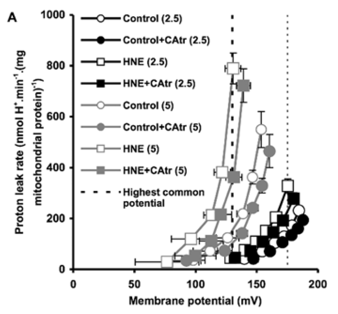
Figure 3 is perhaps the most interesting. For section C they blocked the function of ATP synthase with oligomycin to raise delta psi and then titrated delta psi downwards by uncoupling with FCCP to give either a low or high delta psi. Then they fed the preparation with either palmitoylcarnitine (plus extra carnitine) or glutamate/malate.


The right hand bar graph is derived from the left hand curve and shows that delta psi has some influence but, under both delta psi conditions, there are many more ROS produced under palmitate oxidation than G/M. Delta psi clearly has an effect but substrate also has a marked influence.

That's the convincing part of the paper. There is no insight as to mechanism but my biases assume it will be F:N ratio related. I might have left it there but I can't.

The rest of the results are hugely influenced by this statement from the methods:

"Unless otherwise stated, determinations were made in the presence of oligomycin (3µg/ml) to inhibit ATP synthesis, a condition used in previous studies of the mechanisms of ROS formation in isolated mitochondria and permeabilized muscle fibers (e.g. see Refs. 4, 13, 34, and 38)."

Translation: We blocked ATP synthase because everyone does it.

So pretty well all of the results appear to have been produced during a complete blockade of ATP synthase.

These preparations are always looking at ROS generation when the only dissipation route for membrane potential is some form of uncoupling (UCPs, NNT, various proton assisted co-transporters)

There is no other way to allow O2 consumption under oligomycin. See last post on Brand's review.

This begs the question of how is it possible to feed a mitochondrial preparation supra maximal amounts of NADH substrates (pyruvate/malate) consuming relatively large amounts of oxygen in the absence of any way of dissipating the proton gradient across the inner mitochondrial membrane without ATP synthase being active?

The answer is that there must be some sort of uncoupling going on. The delta psi of 180mV is completely normal but this does not mean that there is no proton leak through the inner mitochondrial membrane. It merely means that the leak (defined by an O2 consumption of 21nmol O/min/mg of mitochondrial protein) is sufficient to avoid raising delta psi to massive levels in the face of a supra maximal supply of pyruvate/malate. The proton leak is also kept low enough not to drop delta psi. This smacks of regulation.

By comparison palmitoylcarnitine at 18micromol/l has less oxygen consumption and supports a lower delta psi, in the region of 145mV.

This is clear in section A of Figure 3.


Both of these values for O2 consumption under oligomycin *have* to be facilitated via uncoupling.

What is also clear is that, with oxygen consumption lower under palmitoylcarnitine, there is less uncoupling than for P/M. 

Has anyone noticed that in Figure 3 there is a flick between 18µM palmitoylcarnitine and 18µM palmitoylcarnitine plus 2mM carnitine between various graphs?

The authors of the paper considered that, with palmitoylcarnitine, the low delta psi combined with low O2 consumption might be due to an inhibitory effect of fatty acid oxidation intermediates on either FAO itself or on ETC function.

Adding 2mM carnitine appears to remove such intermediates. I've not been in to the chemistry but it looks like the carnitine exports them from the mitochondria. There's something about this in the supplementary data. I'm just accepting it happens for today. About which I'm a little cautious.

Adding the extra carnitine makes the oxidation of palmitoylcarnitine look just like supra maximal P/M or G/M.

Here's the oxygen consumption bar chart from supplementary data Figure 3. We're looking at the left hand pair. White bar is palmitoylcarnitine 18µM consuming (as before) 10nmol O/min/mg. Black bar is after the extra carnitine was added. Oxygen consumption is around 25nmol O/min/mg (and delta psi did the same) and is now comparable across the metabolic substrates, black bars:


This increase in O2 consumption means that, under oligomycin, that uncoupling has markedly increased and is directly equivalent to supra maximal NADH sources.

To me this implies that normal fatty acid oxidation (ie without extra carnitine) is a self limiting process. 

If the paper is correct (don't forget they are working with oligomycin blocked preparations) a high delta psi is not a feature of palmitoylcarnitine oxidation.  The oxidation of palmitoylcarnitine suports ROS generation irrespective of delta psi. To make me really happy it would be nice to generate ROS with different fatty acids and look at the effect of the F:N ratio on ROS generation.

There are certain implications to these thoughts. First is that physiology is very keen to keep delta psi in the region of 180mV or lower and applies some degree of uncoupling to achieve this. This appears to be independent of fatty acid induced uncoupling.

This is possibly very important. Any supra maximal supply of substrate in a non-phosphorylating mitochondrial prep (state 4oligomycin) has to have a method to stabilise delta psi at around 180mV. How? Another post there.

Second is that FAO intermediates down regulate the ETC performance directly, limiting delta psi to 145mV from palmitoylcarnitine 18µM unless those FAO intermediates are removed.

Finally, FAO appears to generate ROS moderately independently of delta psi. Certainly palmitoylcarnitine does.


Now for a long-time-ago throwback:

Does everyone recall the Dutch chaps who didn't eat for 60 hours and so rendered their mitochondria "dysfunctional"?

Prolonged fasting identifies skeletal muscle mitochondrial dysfunction as consequence rather than cause of human insulin resistance

Permeablised  muscle fibres behave pretty much like mitochondrial preparations.

"Despite an increase in whole-body fat oxidation, we observed an overall reduction in both coupled state 3 respiration and maximally uncoupled [here using FCCP] respiration in permeabilized skeletal muscle fibers..."

The RCR using an uncoupler and oligomycin, ie state 3FCCP / state 4oligomycin, fell markedly with fasting, hence the term "skeletal muscle mitochondrial dysfunction" in the title.

But 60 hours of fasting cannot possibly destroy your mitochondria. People can pushbike hundreds of kilometres over 5 days without eating anything at all. Their mitochondria work.

I would suspect that this is a fully physiological control system designed to cope, at the mitochondrial level of fatty acid oxidation, with a potentially limitless supply of energy from the fatty acids released from adipocytes under low insulin/insulin signalling conditions.

In this study using permeablised muscle fibres you could probably have reversed the effect completely by treating with 2mM carnitine.

But why would you want to? Apart from gaining insight as to what is normal physiology of course.

Summary: fatty acid oxidation is a self regulating system at the level of beta oxidation rather than at the level of the Krebs Cycle. I suspect that the FAO intermediates will act directly on the electron transport chain.

If this is correct it will provide insight in to other elevated FAO conditions, ie obesity with insulin resistance, where fatty acids should modify (appropriately) ETC function to avoid energetic overload.

I've had suspicions that this has to be the case for a long time. Finally I'm getting to see a little progress.

Peter

Brand insights

In the comments to a previous post met4health provided a link to this paper:

Electron Transport Chain-dependent and -independent Mechanisms of Mitochondrial H2O2 Emission during Long-chain Fatty Acid Oxidation

Before we can go in to it in depth we have to define a few terms. For this we need Brand's excellent review

Assessing mitochondrial dysfunction in cells

which clarifies many of the terms related to mitochondrial function and their variations between experimental set ups.

Perhaps first term we should look at is the Respiratory Control Ratio (RCR). If you feed a quiescent isolated mitochondrial preparation with substrate but no ADP it consumes a small amount of oxygen. If the preparation is given a briefly supramaximal concentration of ADP (in the presence of permanently high phosphate) there will be a marked rise in O2 consumption while the ADP is converted to ATP. This peak O2 consumption represents the maximum respiration under relatively "physiological" conditions. This is state 3 respiration.

If left alone the ADP is fairly quickly used up and O2 consumption will drop back to basal levels. This level is state 4 respiration.

The RCR is state 3 (maximum possible O2 consumption) divided by state 4 (basal O2 consumption). Conceptually this is a marker of how good a mitochondrial preparation is at upping ATP production when needed. High RCR suggests excellent function.

People have modified the routes to these numbers. The first modification is converting the "idling" state 4 to a "stationary" state. This is done with oligomycin, a complete ATP synthase inhibitor. There are reasons this is done but for now we can just accept it. Brand uses the term state 4oligomycin or state 4o.

Next modification, instead of looking at maximum O2 consumption under surplus ADP, is to simply use a chemical uncoupler to probe maximum possible O2 consumption. This is the peak value of state 3 or state 3 uncoupled (state 3u). If you happen to have used FCCP as your uncoupler you might use the term state 3FCCP.

So RCR is simplified to O2 consumption under FCCP divided by O2 consumption under oligomycin. Crude but effective.

Also Brand says this:

"Net forward flux through each electron transport complex requires a thermodynamic disequilibrium, i.e. the free energy available from electron transfer must be greater than that required to pump protons across the membrane against the pmf."

This translates as: if the proton motive force (pmf) is very high then zero electrons will travel down the ETC, none will reach complex IV and zero O2 will be consumed.

Think about that. Without dissipating a high pmf there is no O2 consumption. This suggests that O2 consumption in state 4oligomycin must be synonymous with uncoupling of various types to allow any O2 consumption at all (necessitating pmf dissipation) in the absence of a functional ATP synthase.

This lets you qualify the information provided by papers which quantify the O2 consumption under oligomycin without mentioning the significance of oxygen being consumed in the absence of a functional ATP synthase.

Brand's review is full of such gems as the above but I think these insights are enough to allow us to go on and look at ROS generation from fatty acid oxidation in the presence/absence of elevated pmf (the electrical component of which is delta psi).

Peter

Monday, May 16, 2022

Deuterium protected linoleic acid

This is a fascinating paper which has distracted me from my thought train on uncoupling, mitochondrial membrane potential and ROS generation because it has aspects involving all three while not being particularly intuitive as to what is going on. I picked it up via comments from Tucker and Raphi on twitter.

Deuterium-reinforced polyunsaturated fatty acids protect agains atherosclerosis by lowering lipid peroxidation and hypercholesterolemia

First aside: Mouse model. This current model, the APOE*3-Leiden mouse, is a model. It's not as totally useless as an APOE total knockout or an LDL receptor knockout model but it's still nothing like a real mouse or like a real human. Mouse lipoprotein management is not like human lipoprotein management. They do not have a cholesterol ester transfer protein. The Leiden mouse has this human gene engineered in. It also has a selective APOE*3 knockout to give a mild elevation of LDL. The end result is a model which has numbers on a lipid panel which look a bit more like a human with metabolic syndrome than the average extreme knockout mouse model.

But it's not a human with metabolic syndrome. It's an APOE*3-Leiden mouse and if you found a cure for its "atherosclerosis" you would have a great tool for helping APOE*3-Leiden mice. Would it translate to helping humans with metabolic syndrome? Hahahahahahahahah bonk. End aside.

Second aside: Does LDL cause atherosclerosis? Hahahahahahahah, bonk. To anyone with any sense atherosclerosis is a response to injury where IGF-1 delivered by platelets attaching to the injured endothelium causes media hypertrophy to reinforce the site of injury. This is accelerated if systemic hyperinsulinaemia also acts as an agonist on those IGF-1 receptors. It can almost certainly be enhanced by delivering lipid peroxides such as 4-HNE, 13-HODE and 9-HODE, though their effects are very, very complex. I'm perfectly willing to believe that any genetic engineered tweak in to a mouse which increases the persistence of linoleic acid containing lipoproteins in the plasma allows time for that LA to spontaneously oxidise and accelerate what looks a bit like atherosclerosis, in the model. Just my view. End second aside.


OK. On to the paper:

The APOE*3-Leiden mice were reared on non specific chow. At 12 weeks of age (Time -4) they were put on to something derived from the AIN-93M diet. All lipids were all supplied as methyl esters of fatty acids, not triglycerides. It contained 1.2% of calories as LA and 9% of calories as sucrose. The intervention group had exactly half of the 1.2% of LA calories supplied in the form of deuterium stabilised, ROS peroxidation resistant D2-linoleic acid.

They were fed these diets for four weeks. At that point (Time zero) 0.15% by weight of reagent grade cholesterol was added to both diets (otherwise the model doesn't work to get the essential-for-funding lipid lesions, no sniggering at the back. It's a model). This "western" diet, which only differed from the run-in diet by the added 0.15% reagent grade cholesterol, caused/allowed some weight gain over the following 12 weeks but less in the D2-LA supplemented group than the normal LA group:


No weight gain on either of the run-in diets, followed by lots of gain in the "normal" LA diet but not the D2-LA diet once the cholesterol was added.

Why?

Why should adding 0.15% of cholesterol produce such diverging weight gains?

Even more exciting is if you look at lean mass vs adipose mass:


Adding just 0.15% cholesterol produced a marked fat mass gain in the normal LA mice and a trend downward in fat mass for the ROS protected D2-LA mice.

On top of that the D2-LA mice started eating extra during the period of fat loss. A lot extra:


Soooooooo. What is going on?

Well, the first thing to realise is that during the four week run-in period after the replacement of chow by the AIN-93M derived diets there were already changes which didn't show in total body weights (graphs A and B). The mice, with or without deuterium stabilised LA, all lost muscle mass and all gained fat mass during those weeks, just under a gram of each. So, even without the reagent grade cholesterol, changes were already on going from a "normal" mouse phenotype on chow towards a "skinny-fat" phenotype on an AIN-93M-like diet. That's clear in graphs C and D. Possibly from the sucrose but there's no way of telling that from the paper.

The changes were on-going before the diets were "westernised" by the addition of 0.15% of reagent grade cholesterol. I suspect that the addition of the cholesterol is a red herring.

Let's go on to look at food intakes.

The mice on the deuterated LA eventually began eating more than those on the standard LA. This became statistically significant at about week five.


Any mouse which is eating extra and losing adipose tissue is either showing malabsorption or uncoupling.

I'll buy the uncoupling, but then I would.

Why the delay to the onset of starting to eat extra? Is there a delay in uncoupling onset? Not necessarily. A normal mouse uses a significant percentage of its caloric intake to generate heat in its brown adipose tissue. There is no need to increase food intake while ever the purported uncoupling from deuterated D2-LA is generating less heat than is needed to maintain body temperature. As heat production increases over time it begins to exceed this essential minimum and so comes to represent a "calories-out" in excess of what is merely needed to keep warm. At this point an increase in food intake becomes necessary to balance the heat lost by supra-physiological uncoupling.

If this is correct, and that's a big if, there is clearly an on-going progressive increase in uncoupling with time. The logical explanation is that there is a progressive increase of deuterated D2-LA in tissues and/or being used for beta oxidation.

How might deuterated, ROS resistant LA, facilitate uncoupling?

I don't know, so it's time for some routine wild speculation. If we could answer that one question the whole scenario becomes straight forward. Sadly it is not at all obvious why D2-LA should facilitate uncoupling. Here's my current best shot. If I think of something more plausible I'll post again:

Let's assume that linoleic acid, whether deuterated or native, allows excess calories in to a cell. This is the doodle from a few posts ago:


which then leads to this doodle:


and this doodle:


This begs the question as to how much damage (signalling?) is done by the stray electrons, how much by superoxide, by hydrogen peroxide or how much is actually mediated by the lipid peroxides generated from linoleic acid per se. Which is the most important mediator?

Let me suggest that D2-LA allows the excessive delta psi, which facilitates both reverse electron transport and pathological ROS generation. As in the previous post the high delta psi eventually allows D2-LA to drive RET at the cost of, via high delta psi, allowing electrons to be lost from the ETC to oxygen at abnormal sites, forming superoxide. Under D2-LA this superoxide has very limited ability to contact oxidisable native linoleic acid.

So now we look more like this with ;


There is a surfeit of ATP, high delta psi and availability of either LA and/or D2-LA to facilitate uncoupling combined with minimal damage to the ETC. You do have to have a source of 4-HNE or a related "damage marker" to facilitate uncoupling but it doesn't need much.

I can't see any more straightforward technique for D2-LA to uncouple. If there is one and it's clear how it works, that would be great. Currently this is the best I can do.

Summary: D2-LA allows uncoupling. That explains everything, but the mechanism for the uncoupling is obscure and I'm guessing.

NB I was also trying to explain to myself why the control group got fat. I don't think they did. A total weight gain of 5g over 12 weeks to give a final weight of 25g sounds like a normal mouse to me. It's the slim mice eating extra food to maintain that slim bodyweight that are abnormal.

Ultimately the paper poses the question: What determines whether a cell deals with excess calories by sequestration to storage vs uncoupling. Obviously this is insulin signalling. But is it an oxidation product of linoleic acid which controls insulin signalling? Are we simply looking at a situation of absolutely suppressed insulin signalling, due to D2-LA being "too" stable?

Peter

Final addendum/aside. There is a claim on 'tinternet, un referenced, that low dose oral deuterium oxide in mammals causes weight loss, rather than the death which is the normal result of high dose exposure. Could D2O trigger uncoupling irrespective of LA type and the catabolism of D2-LA be a simple source of deuterated water by oxidation of the D2-LA? I doubt this but the idea is still a potential explanation. I have hunted support/refutation for this without success.