Monday, July 28, 2025

Protons (82) Size matters

Here's the next paper:


I would suggest that the first of many problems with the paper is that it is using ob/ob and db/db mice. I don't know about ob/ob mice but in db/db mice we already know that obesity is only a feature of the genotype when linoleic acid is present in the diet at somewhere above 5% of calories. I discussed Valerie Reeves' PhD here. So the db/db mouse obesity phenoptype is really just a consequence of the poor ROS generation exemplified by linoleic acid. Offsetting this with stearic acid is 100% protective against obesity.

Think about that.

If anyone goes through the paper they will also note that the adipocytes from these mice which exhibit obesity under linoleic acid via low ROS generation are also under extreme oxidative stress, ie high ROS generation, during the process of dying. That's yet another post which will have to wait.
 
On the plus side this suggests that what we see in db/db mice has the potential to be relevant to what happens in D12942 fed bl/6 mice, which the group didn't use for their electron micrographs here. My bias is that db/db mice are remarkably similar to D12942 fed mice in their phenotype. Others may disagree.

So this is a scanning electron micrograph of the degenerating remains of an adipocyte from a db/db mouse:


If anyone wants to go to the image in the paper, it is stunning. You can really, really zoom in.

The large spherical surface is the dying adipocyte. It still has a surface structure of some sort, which is covered in collagen fibrils. See images in Fig 4 for more beautiful microscopy of this. I would suggest that the collagen has been secreted by the macrophages, as they do,

Production of type VI collagen by human macrophages: a new dimension in macrophage functional heterogeneity

to maintain localisation of the large lipid droplet from the dead adipocyte while they deal with it. Okay, okay, here's an histo image from Fig 4, stained for collagen:


and here's a scanning EM of the collagen fibrils on the surface of such an adipocyte, or its remains:


If we go back to a selection of the scanning EM from the top of the post we can see, apparently oozing from the surface, are small lipid droplets (arrows), at least one of which (arrowhead) has been eaten by a macrophage. Each asterisk identifies a macrophage:


We can get an idea of what happens to the lipids in these droplets from here:

Macrophages take up VLDL-sized emulsion particles through caveolae-mediated endocytosis and excrete part of the internalized triglycerides as fatty acids

Well, well, well. Macrophages release of FFAs from the (remains of) adipocytes, a process which, I very much expect, cannot be suppressed by insulin. Would anyone have any expectations as to whether this process, irrespective of any sort of johnny-come-lately-add-on cytokines, might *require* insulin resistance? Evolution builds on what was there already. With modifications.

Where do these FFAs end up if they can't be constrained within adipocytes, for what ever reason? In part they might, if you eat some carbohydrate, be pushed back in to other adipocytes and trigger even more CLS formation, more lipid release, more... Oh wait, week 16 of D12942 feeding looks like this!


Of course the FFAs also end up, to a very large extent, in the liver when adipose cannot mop them up adequately. From the previous CLS paper again:

"However, under conditions of chronic HF feeding, eAT reduction could contribute to lipid overflow to the liver (Fig. 6), potentially exacerbating hepatic insulin resistance (50) or promoting steatohepatitis."

Here's Fig 6:


I look at CLS formation as the end stage of a failure of rising basal lipolysis to limit linoleic acid mediated adipocyte distension. It has the same consequences but more so.

Peter

Thursday, July 17, 2025

Protons (81) Crown like structures

Hyperinsulinaemia, while being unable to suppress basal lipolysis, is still able to facilitate uptake and storage of fatty acids within adipocytes. In this study the rats which were fed an high polyunsaturate diet were fatter and more hyperinsulinaemic than those fed higher saturate diet, all other aspects of the diets being constant.

Diet fat composition alters membrane phospholipid composition, insulin binding, and glucose metabolism in adipocytes from control and diabetic animals


But when you take adipocytes out of the intact rat and ask how well they processed glucose, the high PUFA group adipocytes were *more* insulin sensitive.


The metabolic milieux renders the live rats insulin resistant (elevated FFAs from increased basal lipolysis) while the individual adipocytes extracted from the rats are pathologically insulin sensitive. So the rats become fat and insulin resistant at the macroscopic level but retain insulin sensitivity at the adipocyte level when supplied FFAs are lowered to the tissue culture levels used.

Aside: There is now an on-line calculator which converts ng/ml of insulin to pmol/l insulin. Anyone who has had to convert grams to moles and then get the decimal point correct when converting to picomoles will understand.  Happy happy happy. 100ng/ml is 225pmol/l, a mild post prandial value. 1000ng/ml is 2250pmol/l, aggressive hyperinsulinaemic clamp levels. Most "ordinary" high insulin clamps use the highest post prandial levels of around 1000pmol/l, ie a bit less than half  highest values on the graph above. The above graph is close to physiology. End aside with happy dance.

To me this sets the scene that elevated plasma insulin, combined with enhanced insulin sensitivity at the adipocyte level, can successfully repackage lipids from basal lipolysis back in to adipocytes. That's Carpentier's idea in this paper, as in the last posts.

Increased postprandial nonesterified fatty acid efflux from adipose tissue in prediabetes is offset by enhanced dietary fatty acid adipose trapping

It is also quite possible to break adipocytes by doing this. We have two opposing processes. Enhanced translocation of glucose and fatty acids in to adipocytes under the failure to correctly resist insulin's storage signal, due to inadequate (but far from zero) ROS generation by linoleic acid. The second is the ability of distended adipocytes to release FFAs irrespective of insulin's action by enhanced basal lipolysis. This limits adipocyte size and supplies a competing substrate for insulin sensitive cells which requires the rejection of a certain amounts of glucose (ie insulin resistance) in proportion to the FFAs available from basal lipolysis.

Obviously excess storage, mediated via linoleic acid, wins. Otherwise there would be no linoleic acid mediated obesity. So when an adipocyte is at maximum size and there is a sudden surge in insulin/glucose/FFA availability then the adipocyte will attempt to get bigger. At some point it will fail.

Which leads me on to this D12492 mouse paper:


The photomicrographs are very pretty. The red arrows (placed by the authors) indicate "crown like structures" (CLSs). This is a simple H&E stained image of adipose tissue from a mouse after eating D12492 for eight weeks:


The CLSs appear to be lipid droplets with thickened material surrounding them which looks a lot like cytoplasm. If you go on to use immunohistochemistry to label perilipin A, which labels the protein surrounding the lipid droplet in functional adipocytes, there isn't any. The numbers indicate individual CLSs. Golden brown indicates perilipin A, clearly stained in the (un-numbered) living adipocytes:


If you stain the same section with F4/80, which picks out macrophages, you get this:


which shows that what, on H&E, looks like s thick surround of adipocyte cytoplasm, is in fact a population of macrophages surrounding the remains of a dead adipocyte. Big Eaters. They are clearing up debris

This is the image from a mouse sacrificed after 12 weeks of eating D12942:


and by week 16 we have this


There are no adipocytes visible in this image. It's all crown-like structures. The authors have not placed arrows because they would need to be everywhere. By 16 weeks of the mice eating D12942 their adipose tissue is in crisis, many adipocytes are dead and there is a marked inflammatory response which is clearing up the debris.

By 20 weeks there is significant recovery of adipose architecture, presumably from a supply of stem cells/preadipocytes, but the formation of CLSs continues, a consequence of the continued feeding of D12942. This is the view at week 20 when the study ended:


There is nothing tidy about the death of adipocytes during the formation of CLSs under D12942. The process is known as pyroptosis. I'm not sure how real pyroptosis might be, after all ferroptosis is a well recognised and well researched process which seems to be little more that linoleic acid intoxication. But assuming pyroptosis is real it is considered to be part of the innate immune system by which cells, when they have certain types of overwhelming infection, kill themselves. The process is messy.

Macrophages don't like mess. They get in there to sort it out. They also signal to the rest of the body that something is very wrong and it's time to optimise metabolic conditions to maximally enhance immune function.

Here's what it looks like if you immunostain the macrophages of CLSs for TNF-α or Il-6


Of course both of these cytokines will cause insulin resistance. Which is adaptive (another post there, you think the innate immune system does stupid things?). Not only in the surrounding adipocytes but also systemically. Which leaves a few open questions.

There are thinkers who surmise that adipose tissue inflammation is causal of insulin resistance and even that this insulin resistance, which generates hyperinsulinaemia, might be the actual cause of obesity. I know it sounds strange, but who knows? Everyone is welcome to their opinion.

Or we could hypothesis, as I do, that linoleic acid is the cause of insulin *sensitivity* which enhances insulin's storage signal (without inflammation) to the point where adipocytes die in association with over distension and there is then a massive inflammatory response as a secondary consequence.

There's a lot more to say about unhappy adipocytes and cytokines but I'll leave this post now by suggesting that the residual insulin resistance seen in IGT when FFAs are normalised by acipimox, as in here:


might be mediated by TNF-α, IL-6 and their kindred signaling molecules from CLSs.

Sadly even this may not be quite as simple as it sounds.

Peter

Wednesday, July 16, 2025

Protons (80) The Carpentier Paradox (Carpentier III)

Preamble.

Direct quotes from Carpentier:

"Raglycerol, a marker of total AT lipolytic rate..."

"Plasma glycerol appearance was lower in IGT..."

"Postprandial palmitate appearance (Rapalmitatewas higher in IGT..."

If we combine the second two statements we can re write the findings as:

In people with IGT the rate of lipolysis is decreased (glycerol release) and simultaneously increased (FFA release) in the post prandial period.

A paradox. Oooh exciting! It would have made a great title for the paper.

So I wrote this post.


Just a one liner based on Tucker's link:

Obesity and metabolic perturbations after loss of aquaporin 7, the adipose glycerol transporter


If you knock out the glycerol/water transporter aquaporin 7 you get an obese mouse model, late onset.

This KO increases the glycerol content of adipocytes and, in all probability, drives the reaction

glycerol + ATP <-> glycerol-3-P + ADP

to the right, on the basis of increased glycerol concentration. The enzyme is glycerokinase.

This using a sledge hammer to move reaction kinetics and I doubt it has much to do with generic obesity.

But it does demonstrate that if you drive glycerol-3-phosphate formation you can drive obesity. Then comes this little snippet from the discussion:

"Lazar and coworkers demonstrated that thiazolidinediones markedly increased Gyk [Glycerokinase] mRNA level in adipocytes, resulting in triglyceride accumulation through enhancement of the conversion of glycerol into glycerol-3-P (21)."


The glitazones allow "futile" cycling of FFAs from triglycerides back in to triglycerides WITHOUT releasing the glycerol from the cell. Like aquaporin 7 KO mice but without all of that complicated genetic engineering.

Aside: "Futile" cycling is anathema to evolution. You either have an unavoidable thermogenic effect of an essential process, like protein catabolism, or you have a useful thermic effect like thermogenic uncoupling. The latter is derived from essential uncoupling to avoid damaging elevations of delta psi in mitochondria, wastefull but essential. Futile cycling without fulfilling a need or without an essential underlying process wastes energy which should be used to make babies. Survival of the fecundest is how it goes. "Futile" cycling is pathology. End aside.

So you cannot use glycerol release as an index of total lipolysis if subjects are taking glitazones to become fat. Oops, I mean to become insulin sensitive. Ah, is there any difference?

Which brings us right back to Carpentier's failure to discuss the *fall* in glycerol release from adipocytes concurrent with the *rise* in FFA release in the post prandial period.

Of course Carpentier's subjects weren't taking pharmaceutical activators of PPARγ.

But they were Canadians who had managed to eat sufficient linoleic acid to get themselves in to prediabetes.

Which begs the question: Is linoleic acid a glitazone mimetic? Well, no. But it generates functional PPARγ activators which *are* glitazone mimetics. You know, 9-HODE, 13-HODE and, of course, 4-HNE. All of which, at the correct concentration, would activate PPARγ and allow "futile" cycling of intra-adipocyte FFAs back to triglycerides without releasing their glycerol.

I'm embarrassed that I was unaware of this.

Carpentier is being paid a group leader's salary to be unaware of it. Also, who the hell scrutineered the paper?

Oops. And oops.

Peter

Monday, July 07, 2025

Protons (80) Carpentier II


I've been wanting to write about this paper for some time. But it annoys me. A lot.

Increased postprandial nonesterified fatty acid efflux from adipose tissue in prediabetes is offset by enhanced dietary fatty acid adipose trapping

I only realised yesterday that it is from Carpentier's group. Clearly Carpentier is asking questions about subjects which I am interested in. So it's time to say something.

First comes the title. From my point of view it absolutely concurs with what I would expect. If we accept that people have impaired glucose tolerance because they have accentuated lipid release from adipocytes (due to increased lipid droplet size necessitating elevated basal lipolysis), then storing lipid after a meal *should* increase FFA efflux from adipocytes. Make them big, they then "leak" (in a very controlled manner).

Carpentier used a very comprehensive tracer study to show that this effect is real and does occur (though they didn't look at, and clearly don't have, an hypothetical mechanism). The other finding they report is that this rise in efflux is not from chylomicrons spilling FFAs when they dock with extracellular lipoprotein lipase. The excess FFA efflux comes from adipocyte intracellular lipolysis.

This is consolidated in the first sentence of the abstract:

"The mechanism of increased postprandial nonesterified fatty acid (NEFA) appearance in the circulation in impaired glucose tolerance (IGT) is due to increased adipose tissue lipolysis..."

Both of which confirm my biases. Which makes me want to like the paper.

Here's the fly in the ointment, also from the abstract:

"Plasma glycerol appearance was lower in IGT (P = 0.01), driven down by insulin resistance and increased insulin secretion."

So.

The group is saying that they have documented elevated postprandial FFA efflux from adipocyte lipolysis in subjects with IGT. But they have NOT detected a rise in glycerol from that lipolysis. Quite the opposite.

What's it to be? More lipolysis giving elevated FFA efflux, or less lipolysis giving less glycerol efflux?

You can't have both at the same time. In the abstract and the discussion they are claiming that hyperinsulinaemia secondary to insulin resistance is suppressing glycerol release. But not suppressing (accentuated) FFA release.

Go figure.

So I've sat on the paper, because it confirms most of my biases but doesn't make sense.


The paper is important because, if their FFA flux data are believable, what they are saying is that adipocytes of people with IGT are releasing FFAs in the post prandial period, but there is, at the same time, enhance uptake of FFAs in to adipocytes.

In my terms: accentuated basal lipolysis, which is protective of adipocytes from over distention, is being offset by FFA uptake by adipocytes as a consequence of enhance insulin and insulin signalling secondary to linoleic acid's inability to resist it.

It matters because there is a battle over adipocyte size. When excessive insulin signalling wins over basal lipolysis, people get hurt. Especially their adipocytes do.

The downstream effects are not pretty.

Peter

Saturday, July 05, 2025

Protons (79) Define insulin resistance

It occurred to me while finishing the Carpentier post that it is a beautiful model of metabolic syndrome.

My definition of insulin resistance is an adaptive response to limit insulin-facilitated metabolic substrate ingress in to a cell when an alternative metabolic substrate is being utilised concurrently. With a few caveats.


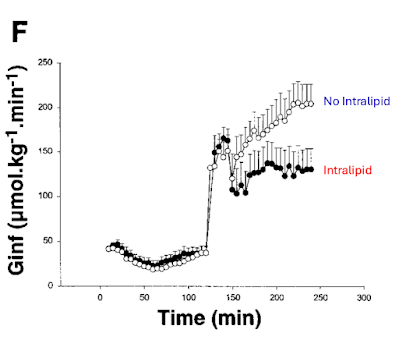
This is exactly what Carpentier generated when he infused Intralipid/heparin to supply FFAs continuously during an hyperglycaemic clamp test. Look at the control group (open circles):


With glucose clamped at 20mmol/l from 120min onward insulin eventually rises to ~700pmol/l which suppresses FFA availability toward the end of the clamp to around 0.050mmol/l or lower. At this point the subjects are running their metabolism almost completely on the glucose supplied by the infusion and FFAs are, appropriately, sequestered in to adipocytes.

The filled circles are the same people but this time, still with glucose clamped at 20mmol/l, they cannot suppress FFAs using insulin because the FFAs are being supplied exogenously using Intralipid. Free fatty acid release from adipocytes will still drop to near zero, as in the control situation, but plasma FFAs are artificially maintained exactly at fasting levels by the infusion.

The insulin resistance of fasting is real. This essential insulin resistance is not some "problem" to be "cured". It is the suppression of glucose uptake when fatty acid generated ROS are signalling that glucose is not needed, so insulin mediated glucose uptake is also not currently needed. Conveniently, this leaves glucose free for use by the brain.

Intralipid here supplies almost exactly the FFAs needed to imitate fasting (~0.70mmol/l) at a time when blood glucose is clamped at 20mmol/l and insulin is high. Resisting insulin under these circumstances is NOT pathology. It is purely adaptive. Fatty acids at 0.70mmol/l supply almost all of a subject's metabolic needs outside of the brain. Subjects do not need the glucose uptake which insulin and hyperglycaemia are trying to force on them. So they resist it. I would do the same.

You can "cure" this insulin "resistance" by turning off the lipid infusion. Probably in less than half an hour, extrapolating from Shulman's work.

So what goes wrong in metabolic syndrome?

The issue in metabolic syndrome is that you cannot turn off the supply of free fatty acids by pressing the stop button on an infusion pump full of Intralipid.

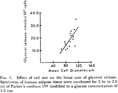
In metabolic syndrome the fatty acids are coming from adipocytes which are larger than they should be and as such have elevated basal lipolysis. We've all read this:

Effect of cell size on lipolysis and antilipolytic action of insulin in human fat cells

showing the effect of cell size on basal lipolysis:


and the inability of insulin, even at preposterous dose rates, to suppress this lipolysis:


So the (inappropriate) fatty acid supply to insulin sensitive cells in obesity *requires* insulin resistance. It is derived from large adipocytes, not small adipocytes (which have low rates of basal lipolysis), ie adipose hypertrophy necessitates insulin resistance while adipose hyperplasia does not. At the same fat mass.

Of course it is possible to stop free fatty acid release mediated through basal lipolysis using acipimox. Again, we've all read this one:

Overnight Lowering of Free Fatty Acids With Acipimox Improves Insulin Resistance and Glucose Tolerance in Obese Diabetic and Nondiabetic Subjects

and struggled to make out the numbers from its spectacularly low quality pdf file illustrations. I think I have the scales correct here:


The insulin tolerance is markedly improved, clearly, but is it not normal. Also, if you work through the rest of the paper, the mitochondria are still far from normal and I have absolutely no problem with adducts of 4-HNE and its relatives causing problems in their own right within the electron transport chain. They are, after all, an intrinsic part of both insulin's activation and deactivation pathways. I wouldn't ignore them. It's a whole series of potential posts about how and why they might be formed. Or not.

But to get back to metabolic syndrome. The obvious question is "Why are adipocytes so big as to be spilling FFAs through size-related elevated basal lipolysis in the first place?".

Insulin. Insulin makes small fat cells in to large fat cells. Stearate is the most effective fatty acid at generating the ROS signal which limits this, with palmitate a close second. Failure to limit insulin signalling, as neatly demonstrated by safflower oil in the Cocoa study, is what makes an adipocyte excessively insulin sensitive and subsequently engorged. With insulin resistance following on as a secondary change derived from the size of adipocytes.

Linoleic acid is a dud for limiting insulin signalling. It's the pathology.

Peter

Thursday, July 03, 2025

Protons (78) Carpentier

I went to Edinburgh for a CPD meeting and skipped social media for four days. I've come back ready to leave it alone for a while longer and to get back to doing some blogging.

Tucker and I have batted this paper, best known as the Carpentier Study, around by email in the past:

Acute enhancement of insulin secretion by FFA in humans is lost with prolonged FFA elevation

and it surfaced in my memory as no tweeted this on X:

"Considering you can use LA to quickly induce IR ... the answer is complicated."

Yes, it's complicated. Both correct and incorrect.

So here is Carpentier's graph of what happens when you use an hyperglycaemic clamp to 20mmol/l, ie the right hand side of the graph where the necessary infusion rate to achieve this concentration is illustrated:


This is completely clear cut. Infusing Intralipid (~50% linoleic acid) for 48h up to and throughout the hyperglycaemic clamp markedly reduces the amount of glucose needed to maintain 20mmol/l in the blood, which signifies insulin resistance.

There are two fundamental problems here. The first is that the subjects were fed, throughout the 48h of the lead up to the clamp, a tightly controlled diet. The total number of calories is not specified but Tucker suggested from other papers by the same group that it was in the region of 2100kcal/d, designed to maintain weight stability.

This was fed either without the Intralipid or with the Intralipid, which provided an additional 1720kcal/24h, if it was included.

So in the "No Intralipid" arm the subjects were on a diet designed to maintain weight stability.

In the "Intralipid" arm the subjects were receiving 3820kcal/d, ie being calorically overloaded during the 48h leading up to the clamp.


Anyone who has even superficially glanced at

Insulin resistance is a cellular antioxidant defense mechanism

will be aware that caloric overload absolutely *should* induce insulin resistance. Otherwise there would be reductive stress (too many calories entering insulin sensitive cells) leading to an excessively high delta psi and subsequent oxidative stress, ie excessive generation of reactive oxygen species. 

The control situation is very different to the Intralipid situation. They are utterly different on a overall calorie supply basis, which is fundamental to the essential adaptive nature of insulin resistance.

Okay.

The second problem (or beauty, next post) is the continuation of the infusion through the hyperglycaemic clamp. In the control situation the subjects were only receiving, intravenously, glucose at the steady state of the hyperglycaemic clamp. Around 200μmol/kg/min.

In the Intralipid arm they were receiving 40ml/h of Intralipid, ie 80kcal/h in addition to the glucose at ~130μmol/kg/min. It's beyond my willpower to convert the Intralipid supply to μmol/kg/min and we don't know the weights of the subjects anyway.

But Protons says that the calories from fat should cause enough insulin resistance to limit insulin facilitated glucose ingress to cells by an amount of calories equivalent to those supplied by the fat. This will happen with Intralipid or any other lipid emulsion, non of which was used, or was available at the time.

The issue Protons has with Intralipid is that it will not cause *enough* superoxide generation, by reverse electron transfer, to adequately resist insulin by the correct amount. If I smooth out the curves from Carpentier's paper we get this:


and if I add in what I would expect an highly saturated fat infusion to produce, we would get this this:


and if I wanted to be perverse I would predict this to be the effect of adding a safflower oil infusion (70% LA) with an even higher linoleic acid content than soybean oil:


Of course this has not been done. What has been done is the Cocoa Study by Xiao (also with Carpentier as co-author) using oral rather than intravenous fats:

Differential effects of monounsaturated, polyunsaturated and saturated fat ingestion on glucose stimulated insulin secretion, sensitivity and clearance in overweight and obese, non-diabetic humans

which again used an hyperglycaemic clamp to 20mmol/l of glucose, which gives exactly what Protons would predict:


The hypothesis that linoleic acid generates insulin resistance promptly, as a direct effect of the generation of reactive aldenhydes formed from linoleic acid in the bloodstream, is not supported by either of the Carpentier papers discussed here.

Far more plausible is the Protons hypothesis in which linoleic acid fails to generate the ROS signal and so fails to correctly limit insulin signalling.

The same ROS signal generates satiety in the brain stem. And it also limits the insulin mediated increase in the size of adipocytes. Linoleate oxidation absolutely causes insulin resistance. No doubt. Unfortunately it doesn't cause enough insulin resistance when compared to the normal physiological mix of palmitate, stearate and oleate.

"It's complicated" applies.

Peter


Luckily the lipid peroxidation hypothesis generates the same message as the Protons hypothesis, limit linoleic acid intake. Maybe it doesn't matter which is correct, excepting it's nice to have an explanation for Carpentier's work. I have to say that the simple message "Linoleate = badness", while beautifully simple, has limited explanatory power for studies like these. "It's complicated" hits the nail on the head.